. 2014 Jan 20;204(2):177-85.
doi: 10.1083/jcb.201306085. Epub 2014 Jan 13.

Motor-driven marginal band coiling promotes cell shape change during platelet activation

Affiliations

Motor-driven marginal band coiling promotes cell shape change during platelet activation

Boubou Diagouraga et al. J Cell Biol. .

Abstract

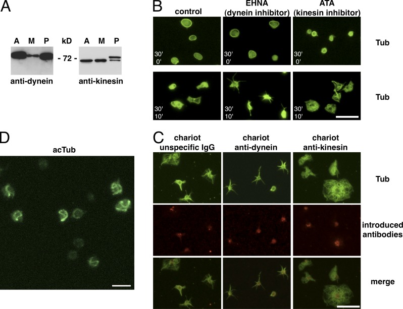
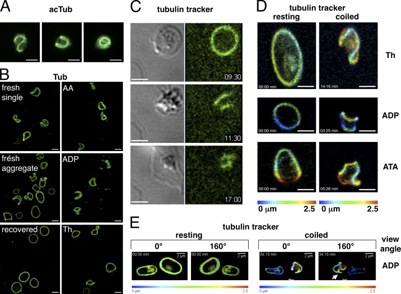
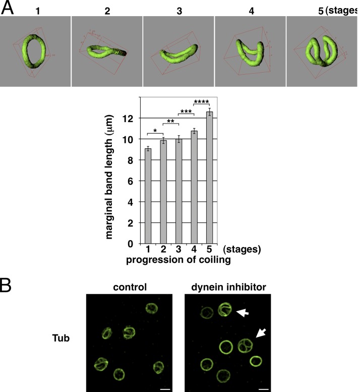
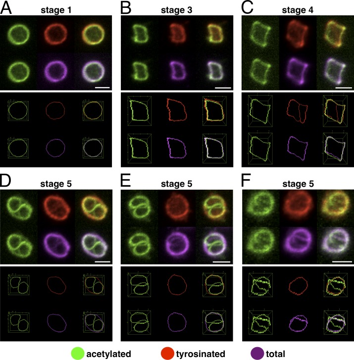
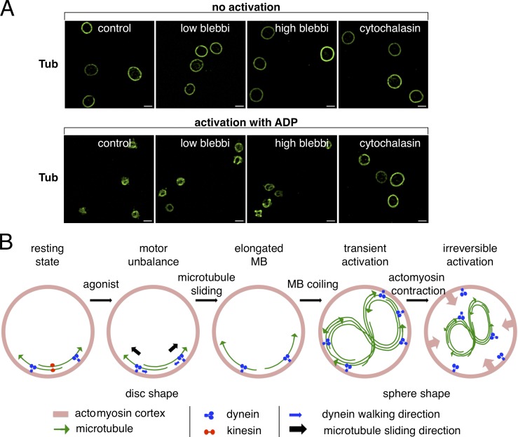
Platelets float in the blood as discoid particles. Their shape is maintained by microtubules organized in a ring structure, the so-called marginal band (MB), in the periphery of resting platelets. Platelets are activated after vessel injury and undergo a major shape change known as disc to sphere transition. It has been suggested that actomyosin tension induces the contraction of the MB to a smaller ring. In this paper, we show that antagonistic microtubule motors keep the MB in its resting state. During platelet activation, dynein slides microtubules apart, leading to MB extension rather than contraction. The MB then starts to coil, thereby inducing the spherical shape of activating platelets. Newly polymerizing microtubules within the coiled MB will then take a new path to form the smaller microtubule ring, in concerted action with actomyosin tension. These results present a new view of the platelet activation mechanism and reveal principal mechanistic features underlying cellular shape changes.


Figures

Figure 1.
Figure 1.
Presence of microtubule motors in platelets and motor inhibitor treatments. (A) Western blot of 5 µg lysates of the cell line A549 (A), the megakaryocyte precursor line CHRF-288-11 (M), and of 107 platelets (P) revealed with a pan-anti–kinesin heavy chain and an anti–dynein intermediate chain antibody. (B) Resting platelets in PRP from buffy coats were diluted in PBS, 2.5 × 10⁶/ml, and incubated with 1 mM EHNA or 10 µM ATA for 30 min at RT and then either fixed (top; 30’ inhibitors/0’ spreading) or allowed to spread on a glass surface for 10 min (bottom; 30’ inhibitors/10’ spreading) before fixation and α-tubulin staining. (C) Control rabbit IgGs as well as mouse anti-dynein and rabbit anti-kinesin function-blocking antibodies were introduced into living platelets using the Chariot kit. Platelets were then allowed to spread on glass coverslips for 10 min, fixed, and stained using a monoclonal rabbit anti α-tubulin antibody for the anti-dynein Chariot and a mouse anti–α-tubulin antibody for the control and the anti-kinesin Chariot conditions (in green) as well as secondary antibodies recognizing the introduced antibodies (anti–mouse for the dynein Chariot and anti–rabbit for the control and the kinesin Chariot conditions, in red). (D) 3D projection of a confocal z stack of platelets treated as in B (top) with 10 µM ATA but for only 3 min, fixed, and stained for acetylated tubulin (acTub). Video 1. Bars: (B and C) 10 µm; (D) 5 µm.
Figure 2.
Figure 2.
MB coiling in activating platelets. (A) 3D reconstruction of confocal z stacks of three representative examples of transiently activated platelets present in PRP prepared from freshly drawn blood, fixed, and stained for acetylated tubulin (AcTub). Video 2. (B, left images) Transiently activated platelets present in PRP prepared from freshly drawn blood: either single platelets, platelets forming a small aggregate, or platelets of the same PRP, which have regained the resting state after a recovery period (120 min at RT). (right images) Platelets present in PRP prepared from buffy coats activated for 60 s with the following agonists: 6.25 µM arachidonic acid (AA), 25 nM ADP, and 0.01 U/ml thrombin (Th). Platelets are stained for α-tubulin. Video 3. (C) Time-lapse video of a microtubule tracker-stained platelet spreading on a glass surface. Transmission (left) and fluorescence (right) images are taken simultaneously every 5 s. Three time points of the activation sequence are shown: disc shape with resting MB, sphere shape with coiled MB, and spread platelet with small microtubule ring. Video 4. Time points are given in minutes and seconds. (D) Two time points of time-lapse videos of 3D reconstructions of confocal stacks of microtubule tracker-stained platelets activated with thrombin (0.017-U/ml final) and ADP (135-nM final) or treated with ATA (6.7-µM final). 3D projections are depth color coded as indicated. Video 5. (E) Two time points of a time-lapse video of 3D reconstructions of confocal stacks of microtubule tracker-stained platelets activated with ADP (270-nM final). 3D projections are depth color coded as indicated. Shown are two different view angles to observe microtubules short cutting the coiled bundle (arrow). Video 6. Bars, 2 µm.
Figure 3.
Figure 3.
Dynein-mediated MB extension induces its coiling. (A) The MB length of resting platelets (PRP prepared from freshly drawn blood after a recovery period) and transiently activated platelets, present in PRP prepared from freshly drawn blood, was measured as described in the Materials and methods section. Platelets were grouped in five categories according to different coiling stages (stage 1, resting platelets, n = 66; stage 2, start of coiling, n = 31; stage 3, slightly coiled, n = 32; stage 4, more coiled, n = 33; and stage 5, maximum coiled, n = 16; data are presented as means ± SEM; *, P < 0.017; **, P < 0.38; ***, P < 0.033; ****, P < 0.0009). (B) Platelets prepared from freshly drawn blood with coiling MBs were diluted in PBS, 2.5 × 10⁶/ml, and incubated with or without EHNA (1-mM final) for 10 min at RT, fixed, and stained for α-tubulin (Tub). Arrows indicate platelets having simultaneously coiled and resting MBs. Video 7. Bars, 2 µm.
Figure 4.
Figure 4.
Newly polymerizing microtubules form a smaller microtubule ring in activated platelets. (A–F) Platelets with MBs at different coiling stages were triple immunostained for acetylated α-tubulin, tyrosinated α-tubulin, and total α-tubulin. Individual stainings as well as merges of 3D reconstructions of confocal z stacks are shown in the top images. The bottom images are generated from the different stainings after thresholding and skeletonizing of the MBs. For further details, see the legends of Videos 8 and 9. Bars, 2 µm.
Figure 5.
Figure 5.
Actomyosin contraction compresses the coiled MB. (A) Platelets were diluted in PBS, 2.5 × 10⁶/ml, and pretreated with or without 25 or 50 µM blebbistatin (blebbi) or 1 µg/ml cytochalasin D for 30 min at RT. Platelets were then either activated with ADP (2.5-µM final) for 10 min at RT (bottom) or not activated (top), fixed, and stained for α-tubulin. Bars, 2 µm. (B) Putative sequence of events leading to platelet activation (to simplify, only two microtubules are shown in disc-shaped platelets).

References

    1. Abramoff M.D., Magalhaes P.J., Ram S.J. 2004. Image Processing with ImageJ. Biophotonics International. 11:36–42
    1. Arganda-Carreras I., Fernández-González R., Muñoz-Barrutia A., Ortiz-De-Solorzano C. 2010. 3D reconstruction of histological sections: Application to mammary gland tissue. Microsc. Res. Tech. 73:1019–1029 10.1002/jemt.20829 - DOI - PubMed
    1. Aslan J.E., Phillips K.G., Healy L.D., Itakura A., Pang J., McCarty O.J. 2013. Histone deacetylase 6-mediated deacetylation of α-tubulin coordinates cytoskeletal and signaling events during platelet activation. Am. J. Physiol. Cell Physiol. 305:C1230–C1239 10.1152/ajpcell.00053.2013 - DOI - PMC - PubMed
    1. Behnke O., Forer A. 1998. From megakaryocytes to platelets: platelet morphogenesis takes place in the bloodstream. Eur. J. Haematol. Suppl. 61:3–23 - PubMed
    1. Bull B.S., Zucker M.B. 1965. Changes in platelet volume produced by temperature, metabolic inhibitors, and aggregating agents. Proc. Soc. Exp. Biol. Med. 120:296–301 10.3181/00379727-120-30516 - DOI - PubMed

Publication types