. 2014 Apr 1;592(7):1677-86.
doi: 10.1113/jphysiol.2013.268573. Epub 2014 Jan 13.

Reflex control of inflammation by sympathetic nerves, not the vagus

Affiliations

Reflex control of inflammation by sympathetic nerves, not the vagus

D Martelli et al. J Physiol. .

Abstract

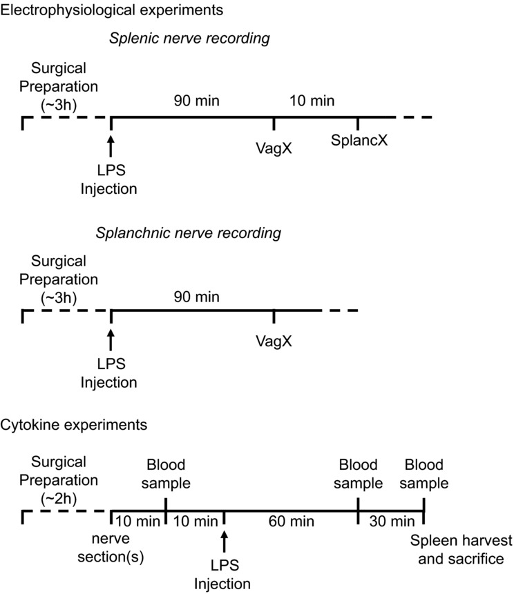
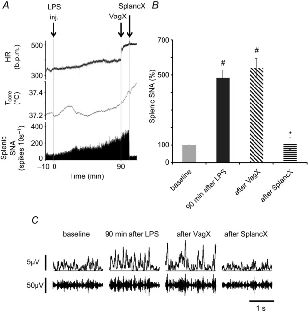
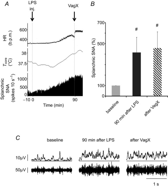
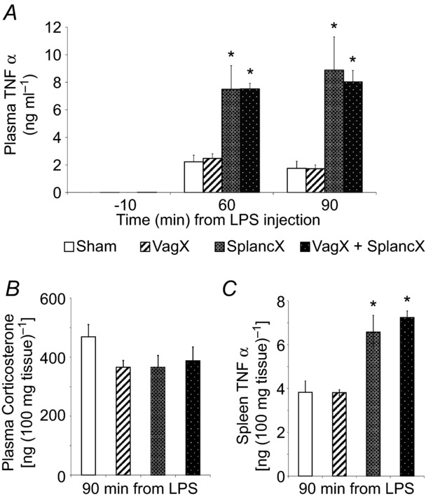
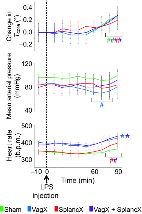
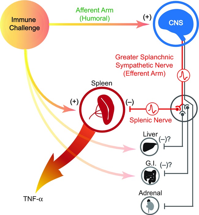
We investigated a neural reflex that controls the strength of inflammatory responses to immune challenge - the inflammatory reflex. In anaesthetized rats challenged with intravenous lipopolysaccharide (LPS, 60 μg kg(-1)), we found strong increases in plasma levels of the key inflammatory mediator tumour necrosis factor α (TNFα) 90 min later. Those levels were unaffected by previous bilateral cervical vagotomy, but were enhanced approximately 5-fold if the greater splanchnic sympathetic nerves had been cut. Sham surgery had no effect, and plasma corticosterone levels were unaffected by nerve sections, so could not explain this result. Electrophysiological recordings demonstrated that efferent neural activity in the splanchnic nerve and its splenic branch was strongly increased by LPS treatment. Splenic nerve activity was dependent on inputs from the splanchnic nerves: vagotomy had no effect on the activity in either nerve. Together, these data demonstrate that immune challenge with this dose of LPS activates a neural reflex that is powerful enough to cause an 80% suppression of the acute systemic inflammatory response. The efferent arm of this reflex is in the splanchnic sympathetic nerves, not the vagi as previously proposed. As with other physiological responses to immune challenge, the afferent pathway is presumptively humoral: the present data show that vagal afferents play no measurable part. Because inflammation sits at the gateway to immune responses, this reflex could play an important role in immune function as well as inflammatory diseases.


Figures

Figure 1
Figure 1
Electrophysiological experiments: surgical preparation and setting up stable nerve recordings took approximately 3 h. Animals were then injected with LPS (60 μg kg−1 i.v.) and followed for 90 min. At that point the cervical vagi were cut bilaterally (VagX). In splenic nerve recording experiments, the ipsilateral (left) splanchnic nerve was cut after a further 10 min (SplancX). Cytokine experiments: after approximately 2 h of surgical preparation, the cervical vagi (VagX), the splanchnic nerves (SplancX), both sets of nerves (VagX + SplancX) or neither (Sham) were severed in those respective groups. After a further 10 min, 1 ml of blood was collected (baseline sample). After a further 10 min, LPS was injected (60 μg kg−1 i.v.). Two further 1 ml blood samples were collected at 60 and 90 min after LPS injection. At 90 min the spleen was also extracted and the rat was killed.
Figure 2
Figure 2
A, example of a chart record showing heart rate (HR, in beats per minute (b.p.m.)), body core temperature (Tcore, °C) and efferent splenic sympathetic nerve activity (SNA) of a urethane-anaesthetized rat injected i.v. with LPS (60 μg kg−1, at ‘inj’). Splenic nerve activity was quantified as spikes per 10 s that exceeded a selected threshold value. Ninety minutes after LPS injection, both cervical vagi were cut (at VagX). Subsequently, the left (ipsilateral) greater splanchnic sympathetic nerve was cut (at SplancX). B, mean splenic SNA in four rats, expressed as a percentage of baseline levels. Statistics were performed on log-transformed absolute values, using a repeated-measures one-way ANOVA followed by a Bonferroni post hoc test. #P < 0.01 compared to baseline levels. *P < 0.01 compared to levels 90 min after LPS. C, examples of raw splenic SNA recordings (lower traces) and rectified, smoothed recordings (10 ms time constant, upper traces) taken at the times indicated. Splenic SNA was significantly increased 90 min after LPS injection, was undiminished by bilateral vagotomy but was significantly reduced by cutting the left greater splanchnic nerve.
Figure 3
Figure 3
A, example of a chart record showing heart rate (HR, in beats per minute (b.p.m.)), body core temperature (Tcore, °C) and efferent activity in the splanchnic sympathetic nerve (SNA) of a urethane-anaesthetized rat injected i.v. with LPS (60 μg kg−1, at ‘inj’). Splanchnic nerve activity was quantified as spikes per 10 s that exceeded a selected threshold value. Ninety minutes after LPS injection, both cervical vagi were cut (at VagX). B, mean splanchnic SNA, expressed as a percentage of baseline levels. Statistics were performed on log-transformed absolute values, using a repeated measures one-way ANOVA followed by a Bonferroni post hoc test. #P < 0.05 compared to baseline levels. C, examples of raw splanchnic SNA recordings (lower traces) and rectified, smoothed recordings (10 ms time constant, upper traces) taken at the times indicated. Splanchnic SNA was increased significantly 90 min after LPS treatment but was undiminished by bilateral vagotomy.
Figure 4
Figure 4
A, plasma TNFα levels in four groups of animals (VagX, SplancX, VagX + SplancX and Sham; each n = 5) in arterial blood samples taken 10 min before, and 60 and 90 min after LPS injection (60 μg kg−1 i.v.). Data were analysed using a repeated-measures two-way ANOVA followed by a Bonferroni post hoc test. *P < 0.001 compared to Sham and VagX levels at the same time point. B, plasma corticosterone levels in the same groups of rats measured in blood samples taken 90 min after LPS administration. No differences were detected between groups (P = 0.194, one-way ANOVA). C, TNFα levels measured in spleens of the same four groups of rats (removed 90 min after LPS injection). *P < 0.01 compared to Sham and VagX treatment (one-way ANOVA followed by Bonferroni post hoc comparison).
Figure 5
Figure 5
Changes of core body temperature, mean arterial pressure and heart rate in response to LPS treatment (60 μg kg−1 i.v.) in four groups of rats, as indicated. Results are expressed as mean ± SEM. Data were analysed using a repeated-measures two-way ANOVA followed by Bonferroni post hoc test. #P < 0.05 compared to baseline levels. *P < 0.05 compared to sham group.
Figure 6
Figure 6
An immune challenge such as lipopolysaccharide (LPS) is detected by cells of the innate immune system, which respond by releasing pro-inflammatory cytokines such as TNFα. In response to systemic LPS, the spleen is known to be the major source of TNFα, which in turn plays a pivotal role in driving the full inflammatory response. At the same time, the immune challenge is detected by the CNS. As in the case of fever, this afferent signal is mediated by humoral factors such as inflammatory cytokines and/or prostaglandins. The CNS then responds by activating a specific subset of sympathetic nerves, whose function is to suppress excessive production of TNFα by the spleen. Other possible targets include the liver, cells in the gastrointestinal (G.I.) tract and the adrenal glands. Those anti-inflammatory nerve fibres run in the greater splanchnic nerve, which constitutes the efferent arm of the inflammatory reflex. Nerve pathways in the vagus do not contribute.

Comment in

References

    1. Andersson U, Tracey KJ. Reflex principles of immunological homeostasis. Ann Rev Immunol. 2012;30:313–335. - PMC - PubMed
    1. Bernik TR, Friedman SG, Ochani M, DiRaimo R, Ulloa L, Yang H, Sudan S, Czura CJ, Ivanova SM, Tracey KJ. Pharmacological stimulation of the cholinergic antiinflammatory pathway. J Exp Med. 2002;195:781–788. - PMC - PubMed
    1. Besedovsky H, del Rey A, Sorkin E, Dinarello CA. Immunoregulatory feedback between interleukin-1 and glucocorticoid hormones. Science. 1986;233:652–654. - PubMed
    1. Borovikova LV, Ivanova S, Zhang M, Yang H, Botchkina GI, Watkins LR, Wang H, Abumrad N, Eaton JW, Tracey KJ. Vagus nerve stimulation attenuates the systemic inflammatory response to endotoxin. Nature. 2000;405:458–462. - PubMed
    1. Bratton BO, Martelli D, McKinley MJ, Trevaks D, Anderson CR, McAllen RM. Neural regulation of inflammation: no neural connection from the vagus to splenic sympathetic neurons. Exp Physiol. 2012;97:1180–1185. - PubMed

Publication types

MeSH terms