Review
. 2014 Apr;34(7):1188-97.
doi: 10.1128/MCB.01454-13. Epub 2014 Jan 13.

The capable ABL: what is its biological function?

Affiliations
Review

The capable ABL: what is its biological function?

Jean Y J Wang. Mol Cell Biol. 2014 Apr.

Abstract

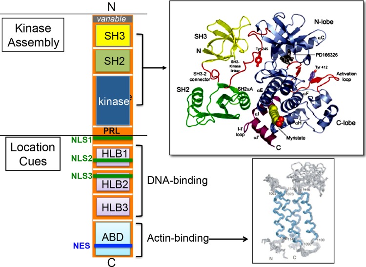
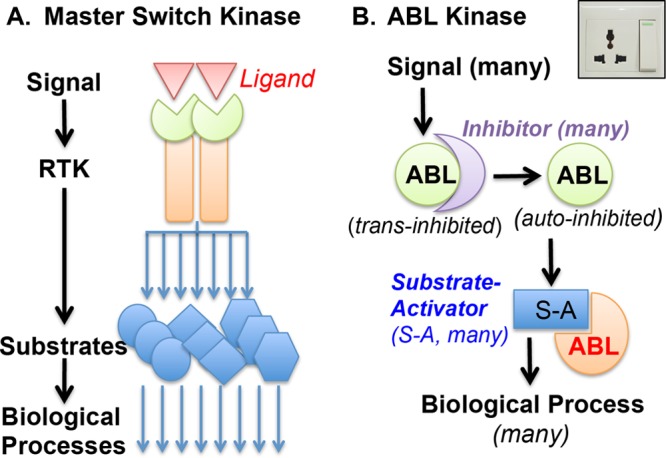
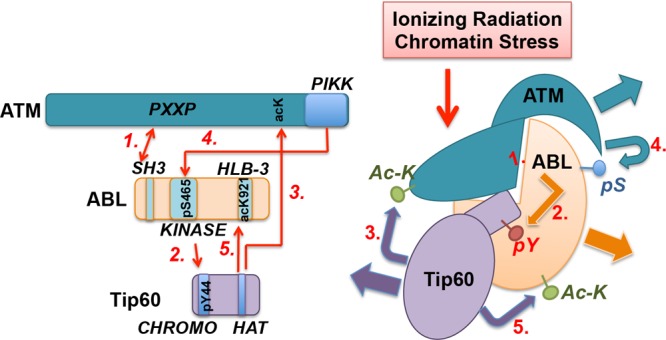
The mammalian ABL1 gene encodes the ubiquitously expressed nonreceptor tyrosine kinase ABL. In response to growth factors, cytokines, cell adhesion, DNA damage, oxidative stress, and other signals, ABL is activated to stimulate cell proliferation or differentiation, survival or death, retraction, or migration. ABL also regulates specialized functions such as antigen receptor signaling in lymphocytes, synapse formation in neurons, and bacterial adhesion to intestinal epithelial cells. Although discovered as the proto-oncogene from which the Abelson leukemia virus derived its Gag-v-Abl oncogene, recent results have linked ABL kinase activation to neuronal degeneration. This body of knowledge on ABL seems confusing because it does not fit the one-gene-one-function paradigm. Without question, ABL capabilities are encoded by its gene sequence and that molecular blueprint designs this kinase to be regulated by subcellular location-dependent interactions with inhibitors and substrate activators. Furthermore, ABL shuttles between the nucleus and the cytoplasm where it binds DNA and actin--two biopolymers with fundamental roles in almost all biological processes. Taken together, the cumulated results from analyses of ABL structure-function, ABL mutant mouse phenotypes, and ABL substrates suggest that this tyrosine kinase does not have its own agenda but that, instead, it has evolved to serve a variety of tissue-specific and context-dependent biological functions.


Figures

FIG 1
FIG 1
Schematic diagram of ABL domains and structures. N, N terminal; variable, alternative promoter and 5′ exon encode two ABL variants, type Ia/b (human) and type I/IV (mouse); SH3, Src homology 3 domain (yellow); SH2, Src homology 2 domain (green); kinase, kinase domain (blue). The crystal structure of the SH3-SH2-kinase assembly with a small molecular inhibitor (PD166316) bound to the kinase N-lobe and a myristate inserted into the kinase C-lobe was solved by Nagar et al. (5). A proline-rich linker (PRL) containing binding sites for the SH3 domains of several substrates such as CRK and NCK is found immediately C terminal to the kinase domain, but this PRL is not included in the crystal structure. NLS, nuclear localization signal; NES, nuclear export signal; HLB, HMG-like box; ABD, actin binding domain. The NMR structure of the ABD has also been solved (6).
FIG 2
FIG 2
ABL is not a master switch kinase. (A) Receptor tyrosine kinase (RTK) is a master switch kinase that is activated by a specific signal—its extracellular ligand—and amplifies that signal by phosphorylating a whole host of intracellular proteins. (B) ABL tyrosine kinase is not a master switch kinase. ABL binds to different trans inhibitors (depicted as a crescent) that partition this nonreceptor tyrosine kinase into different signaling complexes. Signal-regulated release of ABL from a trans inhibitor may not be sufficient for kinase activation if that release does not disrupt the autoinhibitory kinase assembly established by intramolecular inhibitory interactions (Fig. 1) (the autoinhibited ABL is depicted as a full pie in this diagram). In order to become phosphorylated by ABL, a substrate must disrupt autoinhibition. Many ABL substrates identified thus far are ABL-SH3-binding proteins, which disrupt the internal SH3/PXXP interaction to activate ABL (the substrate-bound and activated ABL is depicted as a three-quarter pie in this diagram). Other ABL substrates interact with the SH2 domain or the PRL (Fig. 1), and those interactions may also disrupt autoinhibition. The molecular design of ABL is more similar to an electric socket (upper right) than a master switch (see the text for discussion of this two-step mechanism of restricted ABL activation in signal transduction).
FIG 3
FIG 3
ABL interactions with and modifications by ATM and Tip60 in DNA damage response. This model of ABL interactions and modifications is based on four reports (62–65). Ionizing radiation (IR) or chromatin stress (CS) stimulates ABL interactions with ATM and Tip60. A PXXP motif in ATM binds the ABL-SH3 domain (65) and can thus disrupt the autoinhibitory assembly to activate the ABL kinase (step 1). IR also stimulates ABL interaction with Tip60 (63) and the tyrosine phosphorylation of Tip60 at Tyr44 (62) in the N-terminal CHROMO domain (step 2) by ABL. Tyrosine phosphorylation (pY) of Tip60 stimulates its acetylation of ATM and the activation of ATM kinase (62) (step 3). The activated ATM kinase in turn phosphorylates ABL at Ser465 (pS) (64) (step 4). Ser465 phosphorylation is required for the acetylation of ABL at Lys921 by Tip60 (63) (step 5). Because ABL knockout does not interfere with ATM activation, and because the loss of ABL does not cause radiosensitivity, which is the phenotype of ATM loss (79), ABL is not an obligatory upstream activator of ATM. A simple linear pathway also cannot accommodate the findings that ATM loss causes radiosensitivity whereas ABL loss reduces the apoptosis response but does not cause radiosensitivity. Instead, these interactions are likely to be designed to alter the covalent modifications of ABL (S465 phosphorylation and K921 acetylation) in the DNA damage response.

References

    1. Colicelli J. 2010. ABL tyrosine kinases: evolution of function, regulation, and specificity. Sci. Signal. 3:re6. 10.1126/scisignal.3139re6 - DOI - PMC - PubMed
    1. Greuber EK, Smith-Pearson P, Wang J, Pendergast AM. 2013. Role of ABL family kinases in cancer: from leukaemia to solid tumours. Nat. Rev. Cancer 13:559–571. 10.1038/nrc3563 - DOI - PMC - PubMed
    1. McWhirter JR, Wang JY. 1991. Activation of tyrosinase kinase and microfilament-binding functions of c-abl by bcr sequences in bcr/abl fusion proteins. Mol. Cell. Biol. 11:1553–1565 - PMC - PubMed
    1. McWhirter JR, Wang JY. 1993. An actin-binding function contributes to transformation by the Bcr-Abl oncoprotein of Philadelphia chromosome-positive human leukemias. EMBO J. 12:1533–1546 - PMC - PubMed
    1. Nagar B, Hantschel O, Young MA, Scheffzek K, Veach D, Bornmann W, Clarkson B, Superti-Furga G, Kuriyan J. 2003. Structural basis for the autoinhibition of c-Abl tyrosine kinase. Cell 112:859–871. 10.1016/S0092-8674(03)00194-6 - DOI - PubMed

Publication types

MeSH terms

LinkOut - more resources