. 2014:9:273-87.
doi: 10.2147/IJN.S55118. Epub 2014 Jan 3.

Superparamagnetic iron oxide nanoparticles conjugated with epidermal growth factor (SPION-EGF) for targeting brain tumors

Affiliations

Superparamagnetic iron oxide nanoparticles conjugated with epidermal growth factor (SPION-EGF) for targeting brain tumors

Maxim A Shevtsov et al. Int J Nanomedicine. 2014.

Abstract

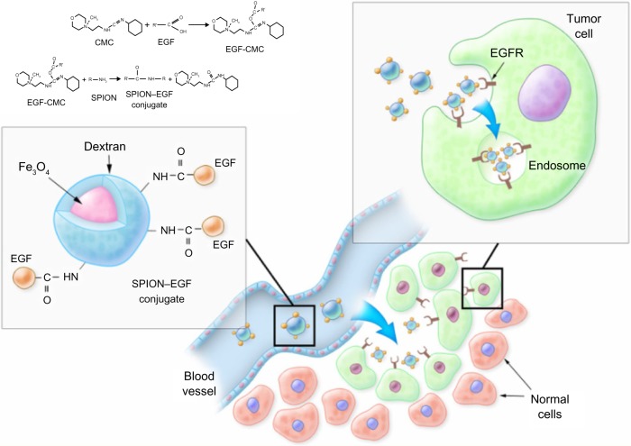
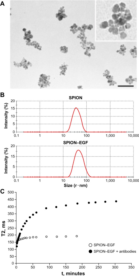
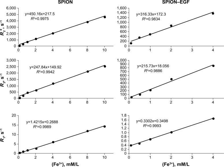
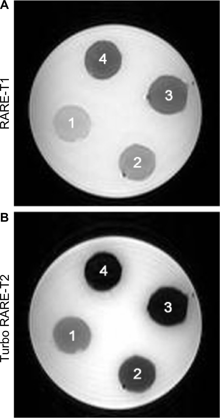
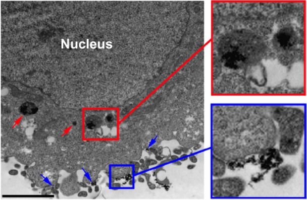
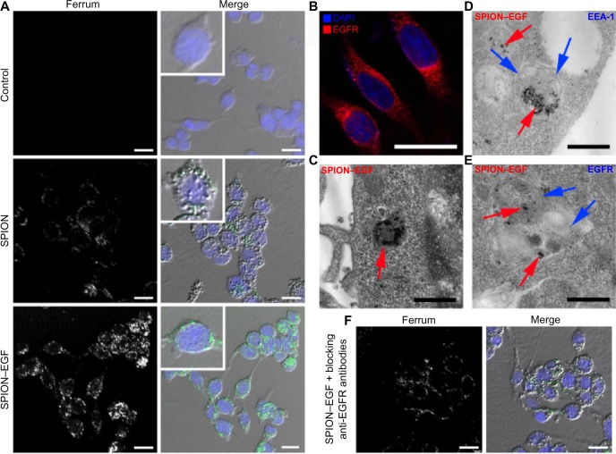
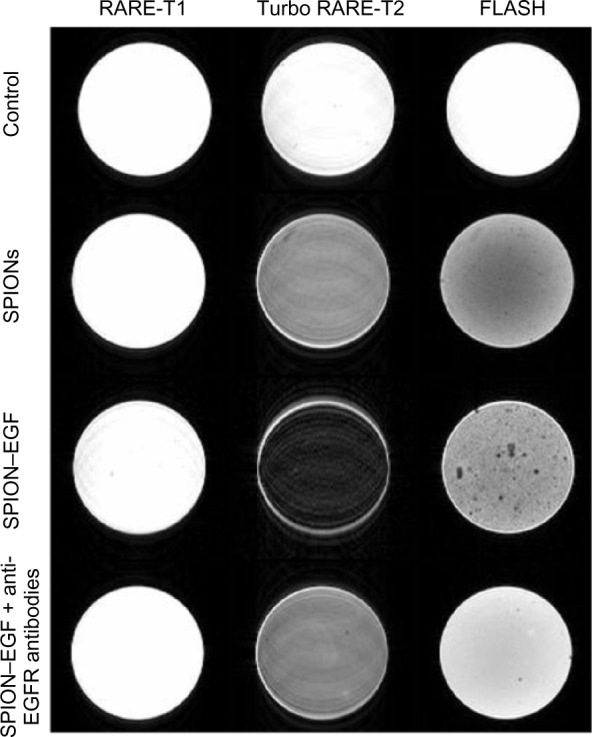
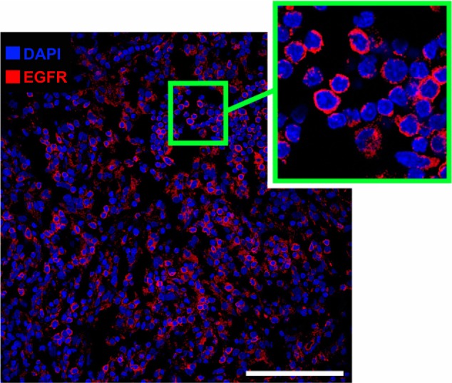
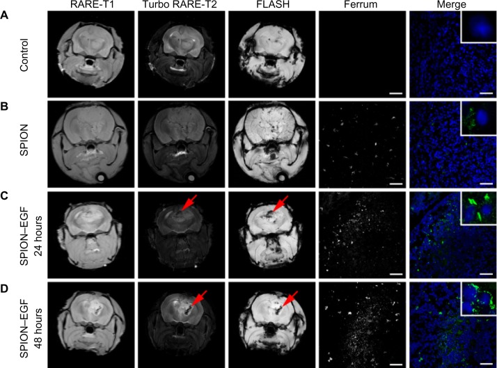
Superparamagnetic iron oxide nanoparticles (SPIONs) conjugated with recombinant human epidermal growth factor (SPION-EGF) were studied as a potential agent for magnetic resonance imaging contrast enhancement of malignant brain tumors. Synthesized conjugates were characterized by transmission electron microscopy, dynamic light scattering, and nuclear magnetic resonance relaxometry. The interaction of SPION-EGF conjugates with cells was analyzed in a C6 glioma cell culture. The distribution of the nanoparticles and their accumulation in tumors were assessed by magnetic resonance imaging in an orthotopic model of C6 gliomas. SPION-EGF nanosuspensions had the properties of a negative contrast agent with high coefficients of relaxation efficiency. In vitro studies of SPION-EGF nanoparticles showed high intracellular incorporation and the absence of a toxic influence on C6 cell viability and proliferation. Intravenous administration of SPION-EGF conjugates in animals provided receptor-mediated targeted delivery across the blood-brain barrier and tumor retention of the nanoparticles; this was more efficient than with unconjugated SPIONs. The accumulation of conjugates in the glioma was revealed as hypotensive zones on T2-weighted images with a twofold reduction in T2 relaxation time in comparison to unconjugated SPIONs (P<0.001). SPION-EGF conjugates provide targeted delivery and efficient magnetic resonance contrast enhancement of EGFR-overexpressing C6 gliomas.

Keywords: C6 glioma; EGFR; MRI contrast agent; SPION; brain tumor; epidermal growth factor; magnetic nanoparticles.


Figures

Figure 1
Figure 1
Scheme of the synthesis and function of SPION–EGF conjugates in tumors. Notes: Synthesis of the magnetic nanoparticle conjugate is presented in the top left. Briefly, dextran-coated nanoparticles were crosslinked with epichlohydrin and were aminated. Activated by CMC, dextran was coupled to carboxyl groups of EGF protein producing SPION–EGF conjugate. Being intravenously injected, nanoparticles (due to the disrupted blood–brain barrier) can accumulate in the tumor site. Receptor-mediated endocytosis of the EGF-functionalized nanoparticles by the EGFR-overexpressing cells (top right) can provide the retention of nanoparticles in the tumor. Abbreviations: CMC, water-soluble carbodiimide; EGFR, epidermal growth factor receptor; SPION, superparamagnetic iron oxide nanoparticle; SPION–EGF, superparamagnetic iron oxide nanoparticles conjugated with epidermal growth factor.
Figure 2
Figure 2
Size and magnetic relaxation assay of the SPION–EGF conjugates in vitro. Notes: (A) Transmission electron microscopy of the SPION–EGF nanoparticles in aqueous dispersion. Scale bar: 100 μ m. (B) Hydrodynamic size (nm) of the SPION or SPION–EGF conjugates in a dilute aqueous dispersion detected by dynamic light scattering using a Zetasizer Nano (Malvern Instruments, Malvern, UK). (C) Magnetic relaxation switch assay on SPION–EGF conjugates. Time evolution of spin–spin relaxation time (T2) of water protons dependent upon the interaction of an EGF conjugate (Fe3+ 0.02 mM/L) with anti-EGF antibodies (0.54 μg/L) in a uniform magnetic field (7.1 T) at 20°C. The magnitude of the change in T2 relaxation time is significantly higher for the sample of SPION–EGF conjugates that interact with antibodies than in the control set without an antibody. Abbreviations: SPION, superparamagnetic iron oxide nanoparticles; SPION–EGF, superparamagnetic iron oxide nanoparticles conjugated with epidermal growth factor.
Figure 3
Figure 3
Magnetic relaxation rates (R2*, R2, and R1) of water protons in a nanoparticle dispersion as a function of the Fe3+ concentration at 20°C. Notes: The inverse magnetic relaxation times (R1 and R2) were calculated from a linear fit of logarithmic echo amplitude versus spin echo time. The values of the magnetic relaxation time observed for water protons in the presence of SPION–EGF conjugates are lower in comparison to non-modified SPIONs due to rapid relaxation of spins in an inhomogenous magnetic field induced by the magnetic nuclei in conjugate. Abbreviations: SPION, superparamagnetic iron oxide nanoparticle; SPION–EGF, superparamagnetic iron oxide nanoparticles conjugated with epidermal growth factor.
Figure 4
Figure 4
Magnetic resonance images of cross sections of agar phantom containing regions with different Fe3+ concentrations of SPION–EGF conjugates. Notes: Presented are the T1 and T2 magnetic resonance images (RARE-T1 and Turbo RARE-T2 regimens, respectively) of the SPION–EGF nanoparticles in 5% agarose gel. 1: 0.1 mM/L; 2: 0.2 mM/L; 3: 0.3 mM/L; 4: 0.4 mM/L. Abbreviations: RARE, rapid acquisition with relaxation enhancement; SPION–EGF, superparamagnetic iron oxide nanoparticles conjugated with epidermal growth factor.
Figure 5
Figure 5
Transmission electron microscopy of the C6 cells incubated with SPIONs. Notes: Following incubation with SPIONs (150 μg/mL) for 24 hours, nanoparticles could be detected as being attached to the cell membrane (blue arrows) and incorporated into the endosome-like structures in the cytoplasm (red arrows). Scale bar: 2 μm. Abbreviation: SPIONs, superparamagnetic iron oxide nanoparticles.
Figure 6
Figure 6
Assessment of the interaction of magnetic nanoparticles with C6 glioma cells by microscopy. Notes: (A) Confocal microscopy images of the C6 cells after 24 hours of incubation with phosphate buffered saline, SPION (150 μg/mL), and SPION–EGF conjugates (150 μg/mL). Nuclei were stained with DAPI (blue). Magnetic nanoparticles were detected by reflected laser scanning at 488 nm (green). Scale bar: 25 μm. (B) Immunofluorescence image of the C6 cells stained by anti-EGFR antibodies (red). Nuclei were stained with DAPI (blue). Scale bar: 7.5 μm. (C) TEM of C6 cells incubated for 24 hours with SPION–EGF conjugates. Electron-dense nanoparticles were present in the cytoplasm of cells in endosome-like structures (red solid arrow). Scale bar: 500 nm. (D) TEM and immunogold labeling of C6 cells incubated with SPION–EGF conjugates for 24 hours. For the detection of early endosomes, a rabbit anti-EEA-1 polyclonal primary antibody and a gold-conjugated (10 nm) goat anti-rabbit IgG secondary antibody were applied. In the cytoplasm of C6 cells, the colocalization of magnetic nanoparticles (red arrows) and the early endosome marker, EEA-1 (blue arrows), was observed in the membrane structures. Scale bar: 500 nm. (E) TEM and immunogold labeling of cells incubated with SPION–EGF nanoparticles. C6 cells were stained with a rabbit polyclonal anti-EGFR antibody and then with a gold-conjugated (10 nm) goat anti-rabbit IgG secondary antibody. The inclusions of nanoparticles (red arrows) were observed in multilayer EGFR-positive endosomes. Scale bar: 500 nm. (F) Confocal microscopy images of the C6 cells treated with blocking anti-EGFR antibodies prior to incubation with SPION–EGF conjugates. A reduction in nanoparticle inclusions in glioma cells (green) was observed. Nuclei were stained with DAPI (blue). Scale bar: 25 μm. Abbreviations: SPION, superparamagnetic iron oxide nanoparticle; SPION–EGF, superparamagnetic iron oxide nanoparticles conjugated with epidermal growth factor; DAPI, 4′,6-diamidino-2-phenylindole; EGFR, epidermal growth factor receptor; EEA-1, early endosome antigen 1; TEM, transmission electron microscopy; IgG, immunoglobulin G.
Figure 7
Figure 7
T1- and T2-weighted, as well as gradient echo magnetic resonance images of C6 cells in agarose gel. Notes: Left column: RARE-T1 regimen; middle column: Turbo RARE-T2 regimen; right column: FLASH regimen. Rows: control cells in phosphate buffered saline; SPION (150 μg/mL); SPION–EGF conjugates (150 μg/mL); cells blocked with anti-EGFR antibodies prior to incubation with SPION–EGF conjugates (150 μg/mL). Abbreviations: RARE, rapid acquisition with relaxation enhancement; Flash, fast low angle shot; SPION, superparamagnetic iron oxide nanoparticle; SPION–EGF, superparamagnetic iron oxide nanoparticle conjugated with epidermal growth factor; EGFR, epidermal growth factor receptor.
Figure 8
Figure 8
Immunofluorescence image of the C6 glioma. Notes: C6 cells were stained with anti-EGFR polyclonal antibodies (red). Nuclei were stained with DAPI (blue). Scale bar: 75 μm. Abbreviations: DAPI, 4′,6-diamidino-2-phenylindole; EGFR, epidermal growth factor receptor.
Figure 9
Figure 9
MR imaging and histology of C6 gliomas in rats. Notes: Coronal MR images in the RARE-T1, Turbo RARE-T2, and FLASH regimes (first three columns). MR scans of (A) control animals (treated with phosphate buffered saline); (B) animals 24 hours after intravenous treatment with SPION (0.3 mg/kg); (C) and animals treated intravenously with SPION–EGF conjugates (0.3 mg/kg) after (C) 24 or (D) 48 hours. Conjugates accumulated in the C6 glioma and presented as hypotense zones on the T2-weighted and gradient echo images (red arrows). Confocal microscopy of the tumor sections is presented in the fourth and fifth columns. Nanoparticles were detected by reflected laser scanning (green); nuclei were stained with DAPI (blue). Scale bar: 75 μm. Abbreviations: RARE, rapid acquisition with relaxation enhancement; FLASH, fast low angle shot; SPION, superparamagnetic iron oxide nanoparticle; SPION–EGF, superparamagnetic iron oxide nanoparticles conjugated with epidermal growth factor receptor; MR, magnetic resonance; DAPI, 4′,6-diamidino-2-phenylindole.

Similar articles

Cited by

References

    1. Wankhede M, Bouras A, Kaluzova M, Hadjipanayis CG. Magnetic nanoparticles: an emerging technology for malignant brain tumor imaging and therapy. Expert Rev Clin Pharmacol. 2012;5(2):173–186. - PMC - PubMed
    1. Hadjipanayis CG, Machaidze R, Kaluzova M, et al. EGFRvIII antibody-conjugated iron oxide nanoparticles for magnetic resonance imaging-guided convection-enhanced delivery and targeted therapy of glioblastoma. Cancer Res. 2010;70(15):6303–6312. - PMC - PubMed
    1. Maier-Hauff K, Ulrich F, Nestler D, et al. Efficacy and safety of intratumoral thermotherapy using magnetic iron-oxide nanoparticles combined with external beam radiotherapy on patients with recurrent glioblastoma multiforme. J Neurooncol. 2011;103(2):317–324. - PMC - PubMed
    1. Tong S, Hou S, Zheng Z, Zhou J, Bao G. Coating optimization of superparamagnetic iron oxide nanoparticles for high T2 relaxivity. Nano Lett. 2010;10(11):4607–4613. - PMC - PubMed
    1. Furnari FB, Fenton T, Bachoo RM, et al. Malignant astrocytic glioma: genetics, biology, and paths to treatment. Genes Dev. 2007;21(21):2683–2710. - PubMed

Publication types

MeSH terms