A different perspective on macroscopic sampling of cholecystectomy specimens
- PMID: 24421844
- PMCID: PMC3887153
- DOI: 10.4132/KoreanJPathol.2013.47.6.519
A different perspective on macroscopic sampling of cholecystectomy specimens
Abstract
Background: Because there may be interdepartmental differences in macroscopic sampling of cholecystectomy specimens, we aimed to investigate differences between the longitudinal sampling technique and our classical sampling technique in cholecystectomy specimens in which there was no obvious malignancy.
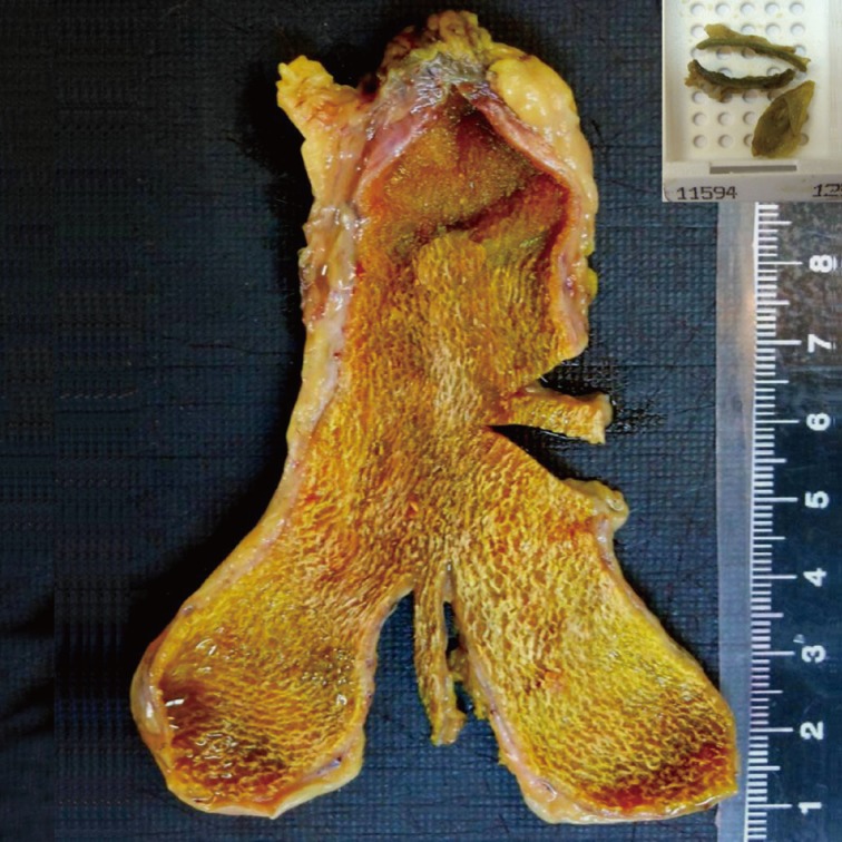
Methods: Six hundred eight cholecystectomy specimens that were collected between 2011 and 2012 were included in this study. The first group included 273 specimens for which one sample was taken from each of the fundus, body, and neck regions (our classical technique). The second group included 335 specimens for which samples taken from the neck region and lengthwise from the fundus toward the neck were placed together in one cassette (longitudinal sampling). The Pearson chi-square, Fisher exact, and ANOVA tests were used and differences were considered significant at p<.05.
Results: In the statistical analysis, although gallbladders in the first group were bigger, the average length of the samples taken in the second group was greater. Inflammatory cells, pyloric metaplasia, intestinal metaplasia, low grade dysplasia, and invasive carcinoma were seen more often in the second group.
Conclusions: In our study, the use of a longitudinal sampling technique enabled us to examine a longer mucosa and to detect more mucosal lesions than did our classical technique. Thus, longitudinal sampling can be an effective technique in detecting preinvasive lesions.
Keywords: Cholecystectomy; Incidental gallbladder cancer; Longitudinal sampling technique; Preinvasive lesions; Swiss-roll method.
Conflict of interest statement
No potential conflict of interest relevant to this article was reported.
Figures


Similar articles
-
Systematic Selective Sampling of Cholecystectomy Specimens Is Adequate to Detect Incidental Gallbladder Adenocarcinoma.Am J Surg Pathol. 2019 Dec;43(12):1668-1673. doi: 10.1097/PAS.0000000000001351. Am J Surg Pathol. 2019. PMID: 31464710
-
Significance of HER2 and Ki-67 in Preneoplastic Lesions and Carcinoma of Gallbladder.J Gastrointest Cancer. 2019 Dec;50(4):848-854. doi: 10.1007/s12029-018-0162-8. J Gastrointest Cancer. 2019. PMID: 30155833
-
[Macro-microscopic comparative study of gallbladder lesions in La Plata (Argentina)].Acta Gastroenterol Latinoam. 1994;24(3):153-8. Acta Gastroenterol Latinoam. 1994. PMID: 7701897 Spanish.
-
Heterotopic gastric mucosa together with intestinal metaplasia and moderate dysplasia in the gall bladder: report of two clinically unusual cases with literature review.Gut. 2001 May;48(5):719-23. doi: 10.1136/gut.48.5.719. Gut. 2001. PMID: 11302975 Free PMC article. Review.
-
Gastric heterotopia together with intestinal metaplasia in the gallbladder: case report and review of literature.Turk J Gastroenterol. 2005 Sep;16(3):160-2. Turk J Gastroenterol. 2005. PMID: 16245229 Review.
Cited by
-
Clinicopathologic Characteristics of Gallbladder Adenomyomas and the Contribution of Macroscopic Sampling in Adenomyoma Diagnosis.Turk Patoloji Derg. 2020;36(1):11-16. doi: 10.5146/tjpath.2019.01471. Turk Patoloji Derg. 2020. PMID: 31633192 Free PMC article.
-
Ultrasonographic features of gallbladder wall thickening in dogs with hypoalbuminemia.Vet Q. 2023 Dec;43(1):1-7. doi: 10.1080/01652176.2023.2240381. Vet Q. 2023. PMID: 37477670 Free PMC article.
-
Immunohistochemical Study of MUC1 and MUC5AC Expression in Gall Bladder Lesions.J Clin Diagn Res. 2017 Jul;11(7):EC12-EC16. doi: 10.7860/JCDR/2017/26537.10230. Epub 2017 Jul 1. J Clin Diagn Res. 2017. PMID: 28892903 Free PMC article.
-
The Contribution of Additional Sampling in Cholecystectomy Materials: A Multicenter Prospective Study.Turk Patoloji Derg. 2020;36(3):188-194. doi: 10.5146/tjpath.2020.01483. Turk Patoloji Derg. 2020. PMID: 32364613 Free PMC article.
-
Differentiating Neoplastic From Non-neoplastic Gallbladder Lesions Using MUC1 and MUC5AC: An Immunohistochemical Analysis.Cureus. 2025 Jul 15;17(7):e88040. doi: 10.7759/cureus.88040. eCollection 2025 Jul. Cureus. 2025. PMID: 40821195 Free PMC article.
References
-
- Mills SE, Carter D, Greenson JK, Reuter VE, Stoler MH. Sternberg's diagnostic surgical pathology. Philadelphia: Lippincott Williams & Wilkins; 2010.
-
- Rosai J. Rosai and Ackerman's surgical pathology. 10th ed. New York: Mosby Elsevier; 2011.
-
- Albores-Saavedra J, Henson DE, Klimstra DS. Tumors of the gallbladder, extrahepatic bile ducts, and ampulla of Vater. Washington, DC: Armed Forces Institute of Pathology; 2000. Atlas of tumor pathology.
-
- Bazoua G, Hamza N, Lazim T. Do we need histology for a normal-looking gallbladder? J Hepatobiliary Pancreat Surg. 2007;14:564–568. - PubMed
-
- Tazuma S, Kajiyama G. Carcinogenesis of malignant lesions of the gall bladder: the impact of chronic inflammation and gallstones. Langenbecks Arch Surg. 2001;386:224–229. - PubMed
LinkOut - more resources
Full Text Sources
Other Literature Sources