

A case of multisystem endometriosis
- PMID: 24421917
- PMCID: PMC3888339
- DOI: 10.3941/jrcr.v7i10.1483
A case of multisystem endometriosis
Abstract
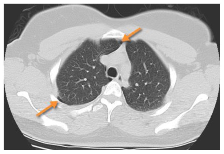
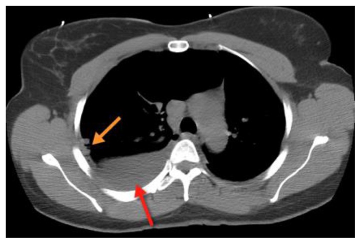
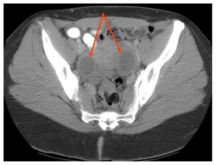
Catamenial pneumothorax is a rare complication secondary to pleural endometriosis. We present a case of a 37-year-old-female with a history of recurrent pneumothoraces with an associated temporal relationship to the onset of her menses. In addition to her recurrent pneumothoraces, on further evaluation, she was found to have multiple nodular masses within the omentum. A thoracoscopic biopsy was subsequently performed, which showed endometrial implants within the pleural space and within the omental cavity. The radiological features and pathogenesis of this rare disease are reviewed and discussed with reference to relevant literature.
Keywords: Endometriosis; abdominal pain; catamenial pneumothorax; chest pain; chocolate cysts; pneumothorax.
Figures

Similar articles
-
A very rare entity: catamenial pneumothorax.Asian Cardiovasc Thorac Ann. 2005 Sep;13(3):271-3. doi: 10.1177/021849230501300317. Asian Cardiovasc Thorac Ann. 2005. PMID: 16113003
-
A Case of Thoracic Endometriosis Syndrome Presenting with Recurrent Catamenial Pneumothorax.Am J Case Rep. 2018 May 17;19:573-576. doi: 10.12659/AJCR.907964. Am J Case Rep. 2018. PMID: 29769481 Free PMC article.
-
Catamenial Pneumothorax: An Unusual Cause of Abdominal Pain.Adv Emerg Nurs J. 2023 Jul-Sep 01;45(3):206-209. doi: 10.1097/TME.0000000000000469. Adv Emerg Nurs J. 2023. PMID: 37501271
-
Catamenial pneumothorax revealing diaphragmatic endometriosis: a case report and revue of literature.Pan Afr Med J. 2017 Jun 14;27:112. doi: 10.11604/pamj.2017.27.112.8007. eCollection 2017. Pan Afr Med J. 2017. PMID: 28819533 Free PMC article. Review.
-
Catamenial pneumothorax: a case report and review of the literature.J Womens Health (Larchmt). 2008 Apr;17(3):367-72. doi: 10.1089/jwh.2007.0553. J Womens Health (Larchmt). 2008. PMID: 18328011 Review.
Cited by
-
Multi-Loculated Catamenial Pneumothorax: A Rare Complication of Thoracic Endometriosis.Cureus. 2021 Aug 30;13(8):e17583. doi: 10.7759/cureus.17583. eCollection 2021 Aug. Cureus. 2021. PMID: 34646637 Free PMC article.
-
Renal Endometriosis Mimicking Cystic Renal Tumor: Case Report and Literature Review.Front Med (Lausanne). 2021 Jun 21;8:684474. doi: 10.3389/fmed.2021.684474. eCollection 2021. Front Med (Lausanne). 2021. PMID: 34235162 Free PMC article.
-
Catamenial pneumothorax due to solitary localization of diaphragmatic endometriosis.Int J Surg Case Rep. 2015;12:19-22. doi: 10.1016/j.ijscr.2015.05.012. Epub 2015 May 7. Int J Surg Case Rep. 2015. PMID: 25981153 Free PMC article.
-
Massive endometrioma presenting with dyspnea and abdominal symptoms.Radiol Case Rep. 2017 Jul 24;12(4):741-745. doi: 10.1016/j.radcr.2017.06.009. eCollection 2017 Dec. Radiol Case Rep. 2017. PMID: 29484061 Free PMC article.
-
Catamenial pneumothorax since introduction of video-assisted thoracoscopic surgery : A systematic review.Wien Klin Wochenschr. 2017 Oct;129(19-20):717-726. doi: 10.1007/s00508-017-1237-4. Epub 2017 Jul 31. Wien Klin Wochenschr. 2017. PMID: 28762057
References
-
- Maurer ER, Schaal JA, Mendez FL. Chronic recurring spontaneous pneumothorax due to endometriosis of the diaphragm. JAMA. 1958;168:2013–4. - PubMed
-
- Marshall MB, Ahmed Z, Kucharczuk JC, Kiser LR, Shrager JB. Catamenial pneumothorax: optimal hormonal and surgical management. Eur J Cardiothoracic Surg. 2005;27:662–666. - PubMed
-
- Alifano M, Trisolini R, Cancellieri A, Regnard JF. Thoracic Endometriosis: Current knowledge. Annals of Thoracic Surgery. 2006;81:761–769. - PubMed
-
- Olive DL, Schwartz LB. Endometriosis. N Engl J Med. 1993;328:1759–69. - PubMed
-
- Joseph J, Sahn SA. Thoracic endometriosis syndrome: new observations from an analysis of 110 cases. Am J Med. 1996;100:164–70. - PubMed
Publication types
MeSH terms
Supplementary concepts
LinkOut - more resources
Full Text Sources
Other Literature Sources
Medical