Review
. 2013 Nov 1;7(11):1-6.
doi: 10.3941/jrcr.v7i11.1817. eCollection 2013 Nov.

Traumatic portacaval shunt: a case report and literature review

Affiliations
Review

Traumatic portacaval shunt: a case report and literature review

Susanna C Spence et al. J Radiol Case Rep. .

Abstract

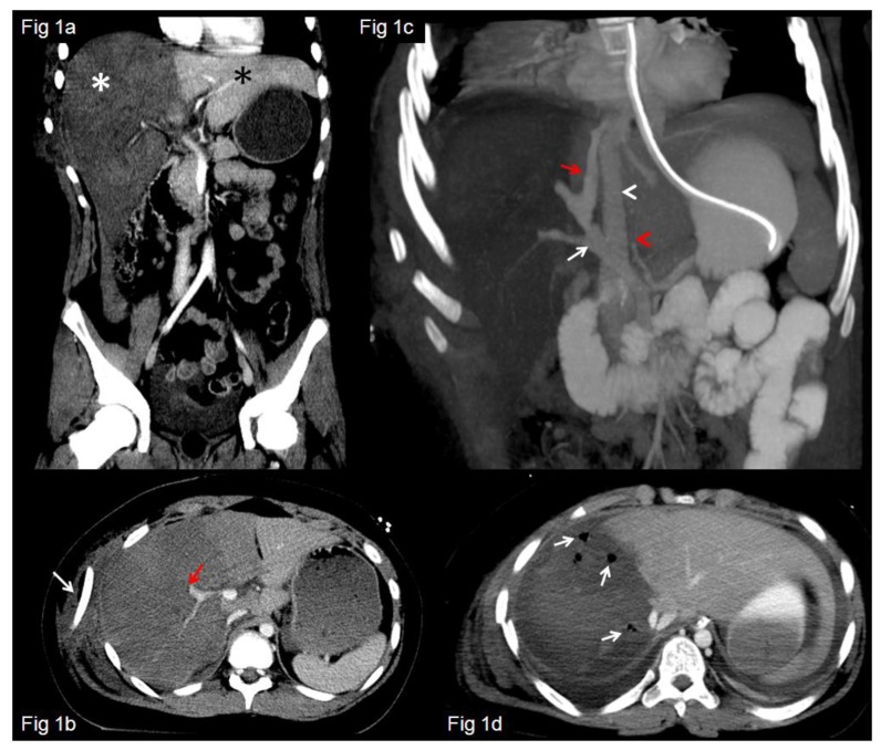
Computed tomography (CT) evaluation of the acute polytrauma patient has become well established as a mainstay of ER triage in hemodynamically stable patients. The radiologist plays a pivotal role in directing management by identifying and appropriately categorizing the severity of a patient's injuries. High-grade liver injuries have undergone an increasing trend of nonoperative management over the last several decades, with concurrent decrease in mortality. However, we present a case of a patient with a grade V liver laceration, in whom a rare portacaval shunt was also present. In the setting of this rare injury, the radiologist will likely be the first person to recognize and categorize a severe complication, which may indicate the need for a fundamental change in patient management.

Keywords: High grade liver injury; grade V liver laceration; traumatic portacaval fistula; traumatic portosystemic fistula; traumatic portosystemic shunt.


Figures

Figure 1
Figure 1
12 year old male with traumatic portacaval fistula. FINDINGS: Coronal CT image through the liver from the patient’s initial trauma CT. Compare the normally perfused and enhancing left lobe of the liver (black asterisk) with the abnormal, devascularized and hypoenhancing right lobe of the liver (white asterisk). Fig 1b: Axial CT image in liver window from the initial trauma CT shows a displaced right 8th rib fracture, which is rotated approximately 180 and displaced laterally (white arrow). There is abrupt occlusion of central branches of the right portal vein (red arrow). TECHNIQUE: Siemens Sensation 40, 5mm slice thickness, 150cc Omnipaque 300, kVp 120, mA 295, coronal and axial plane. Fig 1c. FINDINGS: 2 day follow up CT. Coronal CT image in portal venous phase, through the level of the liver, where an abnormal fistulous connection (red arrow) between the portal vein (white arrow) and suprahepatic IVC (white arrowhead) is well seen, with occlusion of the right portal vein branches distal to the shunt. Abrupt occlusion of the right hepatic artery (red arrowhead) is also evident. Fig 1d: Foci of gas are now identified within the necrotic right lobe of the liver (white arrows). TECHNIQUE: Siemens Sensation 64, multiphase liver CT, kVp 120, mA 345, 5mm slice thickness, coronal and axial plane. Precise contrast type/amount not available.
Figure 2
Figure 2
12 year old male with traumatic portacaval fistula. FINDINGS: 2a and 2b. Multifocal small areas of hypodensity are visualized within the white matter, such as within the left parieto-occipital white matter and left external capsule, suspicious for early edema or ischemic injury. TECHNIQUE: Noncontrast brain CT images, Siemens Sensation 64, 4.8mm slice thickness, kvp 120, mA 450, axial plane.
Figure 3
Figure 3
12 year old male with traumatic portacaval fistula. FINDINGS: Loss of gray-white differentiation, sulcal and ventricular effacement have developed in the interim, consistent with diffuse cerebral edema (white asterisk). Geographic hypodensity within the left thalamus is consistent with an acute left thalamic infarct (white arrow). TECHNIQUE: Noncontrast brain CT images, Siemens Sensation 64, 4.8mm slice thickness, kvp 120, mA 450, axial plane.
Figure 4
Figure 4
12 year old male with traumatic portacaval fistula. FINDINGS: Absence of intracranial perfusion on flow images, and absence of Tc99m ECD metabolism on the delayed images, supporting the clinical diagnosis of brain death. TECHNIQUE: Following IV injection of 25.9mCi Tc99m ECD, dynamic flow images were obtained in anterior projection, followed by 20 minute delayed images in the anterior and lateral projections.

References

    1. Moore EE, Cogbill TH, Jurkovich GJ, Shackford SR, Malangoni MA, Champion HR. Organ Injury Scaling: Spleen and Liver (1994 Revision) J Trauma. 1995;38:323–4. - PubMed
    1. Kozar RA, Moore JB, Niles SE, Holcomb JB, Moore EE, Cothren CC, Hartwell E, Moore FA. Complications of Nonoperative Management of High-Grade Blunt Hepatic Injuries. J Trauma. 2005;59:1066–1071. - PubMed
    1. Ahn JH, Yu JS, Hwang SH, Chung JJ, Kim JH, Kim KW. Nontumorous arterioportal shunts in the liver: CT and MRI findings considering mechanisms and fate. Eur Radiol. 2010;20:385–394. - PubMed
    1. Guzman EA, McCahill LE, Rogers FB. Arterioportal fistulas: introduction of a novel classification with therapeutic implications. J Gastrointest Surg. 2006;10:543–550. - PubMed
    1. Bosoi CR, Rose CF. Brain edema in acute liver failure and chronic liver disease: Similarities and differences. Neurochem Int. 2013 Mar;62(4):446–57. - PubMed

LinkOut - more resources