Case Reports
. 2013 Nov 1;7(11):7-14.
doi: 10.3941/jrcr.v7i11.1559. eCollection 2013 Nov.

Dysembryoplastic neuroepithelial tumor with atypical presentation: MRI and diffusion tensor characteristics

Affiliations
Case Reports

Dysembryoplastic neuroepithelial tumor with atypical presentation: MRI and diffusion tensor characteristics

Kalyan Paudel et al. J Radiol Case Rep. .

Abstract

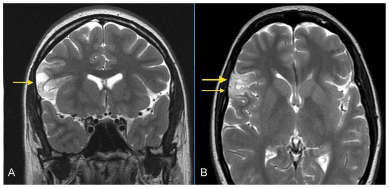
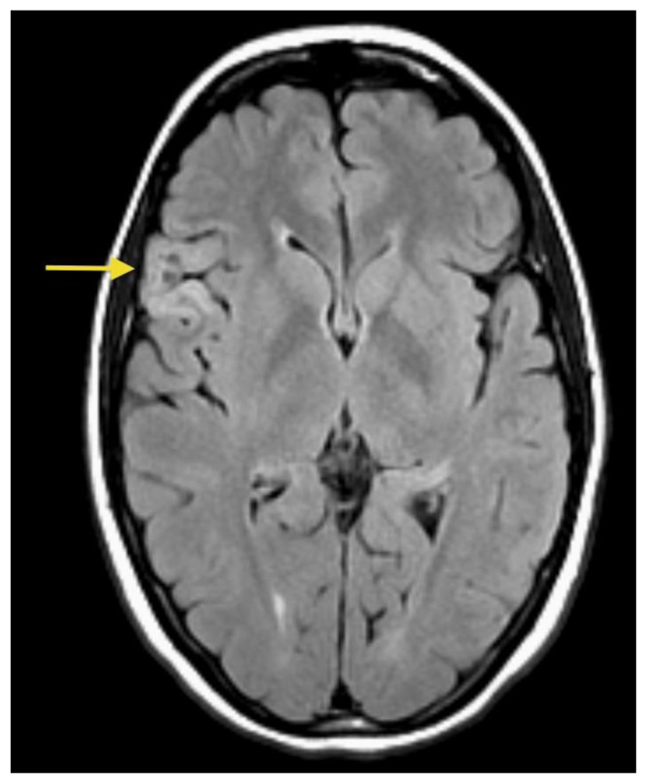
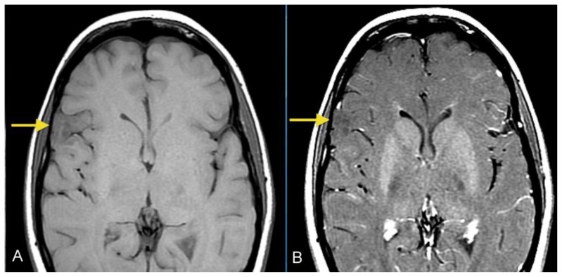
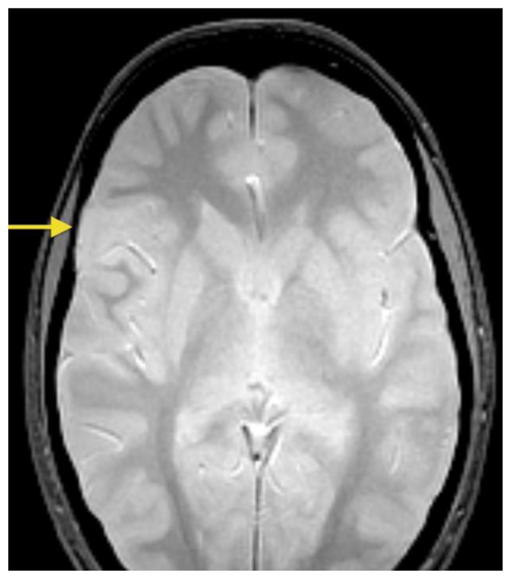
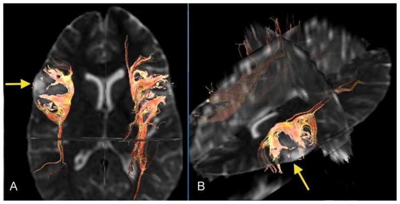
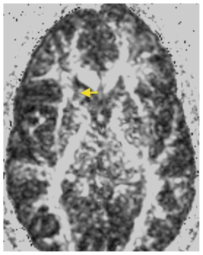
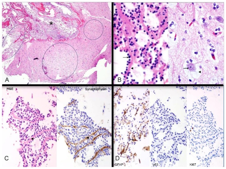
We report the neuroimaging findings of a 26-year-old female patient with a biopsy-proven dysembryoplastic neuroepithelial tumor (DNET). DNETs are an uncommon, usually benign, glial-neural cortical neoplasm of children and young adults who typically present with intractable seizures. DNETs may occur in any region of the supratentorial cortex, but have a predilection for the temporal lobes. Accurate neuroimaging diagnosis is essential since patients with DNET benefit from complete resection. However, accurate differentiation from other cortical lesions may be challenging. Typical conventional Magnetic Resonance Imaging (MRI) features can help in the differentiation from other similar cortical tumors. Diffusion tensor imaging can also provide important additional diagnostic information regarding the degree of involvement of adjacent parenchyma and white matter tracts. In this case, tractography and fractional anisotropy maps demonstrated that fiber tracts surrounding the lesion were displaced, but fiber integrity was maintained, which is more suggestive of a DNET rather than a more aggressive neoplasm. Accurate identification of DNETs is essential for the purpose of rendering a timely diagnosis and start appropriate treatment.

Keywords: Diffusion tensor imaging; Dysembryoplastic neuroepithelial tumor (DNET); Neuroimaging; Tractography.


Figures

Figure 1
Figure 1
26- year-old female with a dysembryoplastic neuroepithelial tumor (DNET) in the right frontal lobe. A: Coronal T2-weighted MR image shows a cortical-based superficial lesion in right frontal lobe extending to the cortical margin. There are multiple small rounded internal focal regions of high signal intensity compatible with a “soap bubble” appearance (arrow). There is no evidence of associated mass effect or vasogenic edema. (Protocol: 1.5 Tesla MRI (GE system), Repetition time/Echo time TR/TE: 3750/76.9, 3mm slice thickness, non-contrast, field of view 24 cm). B: Axial T2-weighted MR image demonstrates superficial lesion in right frontal lobe extending to cortical margin. There are internal focal regions of high signal intensity, compatible with a “soap bubble” appearance (thin arrow). There is also narrowing and remodeling of the adjacent inner table of the skull, compatible with slow growth (wide arrow). (Protocol: 1.5 Tesla MRI (GE system), TR/TE: 3200/256, 5mm slice thickness, field of view 24 cm, non-contrast).
Figure 2
Figure 2
26- year-old female with a dysembryoplastic neuroepithelial tumor (DNET) in the right frontal lobe. Axial FLAIR image show a slightly hyperintense cortex and slightly decreased signal in the small focal nodular areas (arrow). (Protocol: 1.5 Tesla MRI (GE system), TR/TE: 8002/123, 5mm slice thickness, field of view 24 cm, non-contrast).
Figure 3
Figure 3
26- year-old female with a dysembryoplastic neuroepithelial tumor (DNET) in the right frontal lobe. Axial pre (A) and post-contrast (B) T1-weighted images image show a slightly hypointense cortex in the right lateral frontal lobe with no definite enhancement (Protocol: 1.5 Tesla MRI (GE system), TR/TE: 767/16, 5mm slice thickness, field of view 24 cm).
Figure 4
Figure 4
26- year-old female with a dysembryoplastic neuroepithelial tumor (DNET) in the right frontal lobe. Axial gradient echo image shows no signal loss or ‘blooming’ within the mass, indicating no evidence of calcifications or hemorrhage within the mass (arrow). (Protocol: 1.5 Tesla MRI (GE system), TR/TE: 500/9, 5mm slice thickness, field of view 24 cm, non-contrast).
Figure 5
Figure 5
26- year-old female with a dysembryoplastic neuroepithelial tumor (DNET) in the right frontal lobe. Axial Diffusion tensor imaging (DTI) with axial (A) and oblique (B) 3-dimensional images demonstrates deformed and displaced but intact fiber tracts curving around and surrounding the lesion (arrows). The red color is present for visual demonstration of the tracts. (Protocol: DTI was processed with GE Functools software after acquiring data from 1.5 Tesla MRI, GE system, TR/TE: 8500/91.8, 5 mm slice thickness, field of view 24 cm, b-value 1000 s/mm2, number of directions 25)
Figure 6
Figure 6
26- year-old female with a dysembryoplastic neuroepithelial tumor (DNET) in the right frontal lobe. Axial Fractional Anisotropy (FA) map corresponding to Figure 1B shows that the signal in the white matter surrounding the lesion is preserved, suggesting intact fiber integrity (arrow). (Protocol: GE system 1.5 Tesla MRI, TR/TE: 8500/91.8, 5 mm slice thickness, field of view 24 cm, b-value 1000 s/mm2, number of directions 25).
Figure 7
Figure 7
26- year-old female with a dysembryoplastic neuroepithelial tumor (DNET) in the right frontal lobe. Histochemical and immunohistochemical stains of a dysembryoplastic neuroepithelial tumor (DNET) in a 26-year-old male. A: Haematoxylin- and eosin-stained section through the tumor demonstrates discrete intracortical tumor nodules (ovals); the nodule on the left extends into subarachnoid space (asterisk). Hematoxylin and eosin (H&E), high power (40×). B: Histologic features. LEFT: Oligodendroglia-like cells (arrow) lined up along neuropil bundles (arrowhead). RIGHT: Cortical neurons (arrow) floating in myxoid pools (arrowhead). These two features comprise the “specific glioneuronal element” of dysembryoplastic neuroepithelial tumor. Both images H&E, high power (400×). C: Axonal nature of neuropil bundles. The neuropil bundles (arrowheads) in the specific glioneuronal element are immunopositive (brown) for synaptophysin, supporting their axonal nature. LEFT: Hematoxylin and eosin (H& E), high power (200×); RIGHT: synaptophysin immunostain, high power (200×). D: Additional immunohistochemical findings. LEFT: GFAP-immunopositive reactive astrocytes (arrow) are scattered in the specific glioneuronal element. CENTER: Mutant p53 protein is not expressed in the nuclei of oligodendroglia-like cells. RIGHT: Neoplastic cells show a low proliferation index with Ki-67 immunostain. Left: Hematoxylin and eosin (H&E), glial fibrillary acidic protein (GFAP) immunostain, 200×; Center: p53 protein immunostain, 200×; Right; Ki-67 immunostain, 200

Similar articles

Cited by

References

    1. Daumas-Duport C, Pietsch T, Lantos PL. Dysembryoplastic neuroepithelialtumour. In: Kleihues P, Cavenee WK, editors. Pathology and genetics of tumours of the nervous system. Lyon, France: IARC; 2000. pp. 103–106.
    1. Cervera-Pierot P, Varlet P, Chodkiewicz JP, Daumas-Duport C. Dysembryoplastic neuroepithelial tumors located in the caudate nucleus area: report of four cases. Neurosurgery. 1997 May;40(5):1065–1070. - PubMed
    1. Frater JL, Prayson RA, Morris HH, Bingaman WE. Surgical pathological findings of extratemporal-based intractable epilepsy: a study of 133 consecutive resections. Arch Pathol Lab Med. 2000 Apr;124:545–549. - PubMed
    1. Kuroiwa T, Kishikawa T, Kato A, Ueno M, Kudo S, Tabuci K. Dysembryoplastic neuroepithelial tumors: MR findings. J Comput Assist Tomogr. 1994 May-Jun;18(3):352–356. - PubMed
    1. Parmar HA, Hawkins C, Ozelame R, Chuang S, Rutka J, Blaser S. Fluid-attenuated inversion recovery ring sign as a marker of dysembryoplastic neuroepithelial tumors. J Comput Assist Tomogr. 2007 May-Jun;31(3):348–353. - PubMed

Publication types