

Isolated duodenal duplication cyst presenting as a complex solid and cystic mass in the upper abdomen
- PMID: 24421928
- PMCID: PMC3888335
- DOI: 10.3941/jrcr.v7i11.1785
Isolated duodenal duplication cyst presenting as a complex solid and cystic mass in the upper abdomen
Abstract
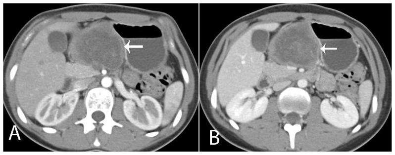
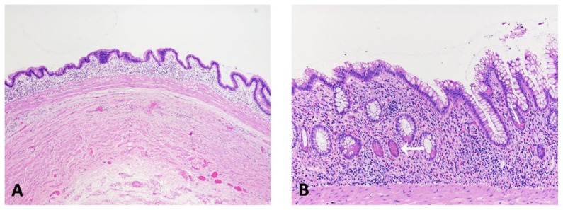
Duodenal duplication cysts are a rare subtype of gastrointestinal duplications cysts. Approximately 5% of gastrointestinal duplication cysts occur in the duodenum. An 18-year-old woman presented with epigastric pain and a subjective abdominal bulge. A computed tomography scan was subsequently performed and showed a solid and cystic mass with wall calcifications in the lesser sac of the upper abdomen. A duodenal duplication cyst was found unexpectedly on histopathologic analysis. This was also an unusual case as there was no evidence of malignancy. Four years after surgery, the patient remains asymptomatic. We present a brief literature review on duodenal duplication cysts and discuss its differential diagnosis.
Keywords: Computed tomography; Duodenum; Duplication cyst; Gastrointestinal stromal tumor; Lesser sac.
Figures

Similar articles
-
Endoscopic partial resection of a duodenal duplication cyst.Endoscopy. 2001 Sep;33(9):808-10. doi: 10.1055/s-2001-16528. Endoscopy. 2001. PMID: 11558037
-
Endoscopic treatment of periampullary duodenal duplication cyts in an 18-month-old girl.Turk J Pediatr. 2023;65(2):344-349. doi: 10.24953/turkjped.2022.1068. Turk J Pediatr. 2023. PMID: 37114701
-
Prenatal sonographic findings of duodenal duplication: case report.J Clin Ultrasound. 2013 Nov-Dec;41 Suppl 1:1-5. doi: 10.1002/jcu.22007. Epub 2012 Nov 2. J Clin Ultrasound. 2013. PMID: 23124691
-
Recurrent acute pancreatitis secondary to a duodenal duplication cyst in an adult. A case report and literature review.Can J Gastroenterol. 2009 Nov;23(11):749-52. doi: 10.1155/2009/979431. Can J Gastroenterol. 2009. PMID: 19893770 Free PMC article. Review.
-
Meta-analysis: the clinical features of the duodenal duplication cyst.J Pediatr Surg. 2010 Aug;45(8):1598-606. doi: 10.1016/j.jpedsurg.2010.01.010. J Pediatr Surg. 2010. PMID: 20713206 Review.
Cited by
-
Duodenal Duplication Cyst: A Rare Differential Diagnosis in a Neonate with Bilious Vomiting.European J Pediatr Surg Rep. 2015 Dec;3(2):82-4. doi: 10.1055/s-0035-1558827. Epub 2015 Sep 10. European J Pediatr Surg Rep. 2015. PMID: 26788454 Free PMC article.
-
Type B choledochocele vs duodenal duplication cyst: a diagnostic dilemma and its management: a case report.J Med Case Rep. 2019 May 24;13(1):160. doi: 10.1186/s13256-019-2010-2. J Med Case Rep. 2019. PMID: 31122272 Free PMC article.
-
Small Bowel Congenital Anomalies: a Review and Update.Curr Gastroenterol Rep. 2016 Apr;18(4):16. doi: 10.1007/s11894-016-0490-4. Curr Gastroenterol Rep. 2016. PMID: 26951229 Review.
-
54-cm enteric duplication cyst in a 13-year-old female.Clin Case Rep. 2018 Sep 21;6(11):2099-2102. doi: 10.1002/ccr3.1751. eCollection 2018 Nov. Clin Case Rep. 2018. PMID: 30455900 Free PMC article.
-
A case of completely isolated advanced enteric duplication cyst cancer performed partial pancreatectomy.Int J Surg Case Rep. 2019;54:83-86. doi: 10.1016/j.ijscr.2018.11.060. Epub 2018 Nov 27. Int J Surg Case Rep. 2019. PMID: 30553095 Free PMC article.
References
-
- Prasad TR, Tan CE. Duodenal duplication cyst communicating with an aberrant pancreatic duct. Pediatr Surg Int. 2005;21:320–322. - PubMed
-
- Hata H, Hiraoka N, Ojima H, et al. Carcinoid tumor arising in a duplication cyst of the duodenum. Pathol Int. 2006;56:272–278. - PubMed
-
- Chen JJ, Lee HC, Yeung CY, et al. Meta-analysis: the clinical features of the duodenal duplication cyst. J Pediatr Surg. 2010;45:1598–1606. - PubMed
-
- Macpherson RI. Gastrointestinal tract duplications: clinical, pathologic, etiologic, and radiologic considerations. Radiographics. 1993;13(5):1063–1080. - PubMed
Publication types
MeSH terms
LinkOut - more resources
Full Text Sources
Other Literature Sources
Medical
Molecular Biology Databases