

Baló's concentric sclerosis: imaging findings and pathological correlation
- PMID: 24421937
- PMCID: PMC3888114
- DOI: 10.3941/jrcr.v7i6.1251
Baló's concentric sclerosis: imaging findings and pathological correlation
Abstract
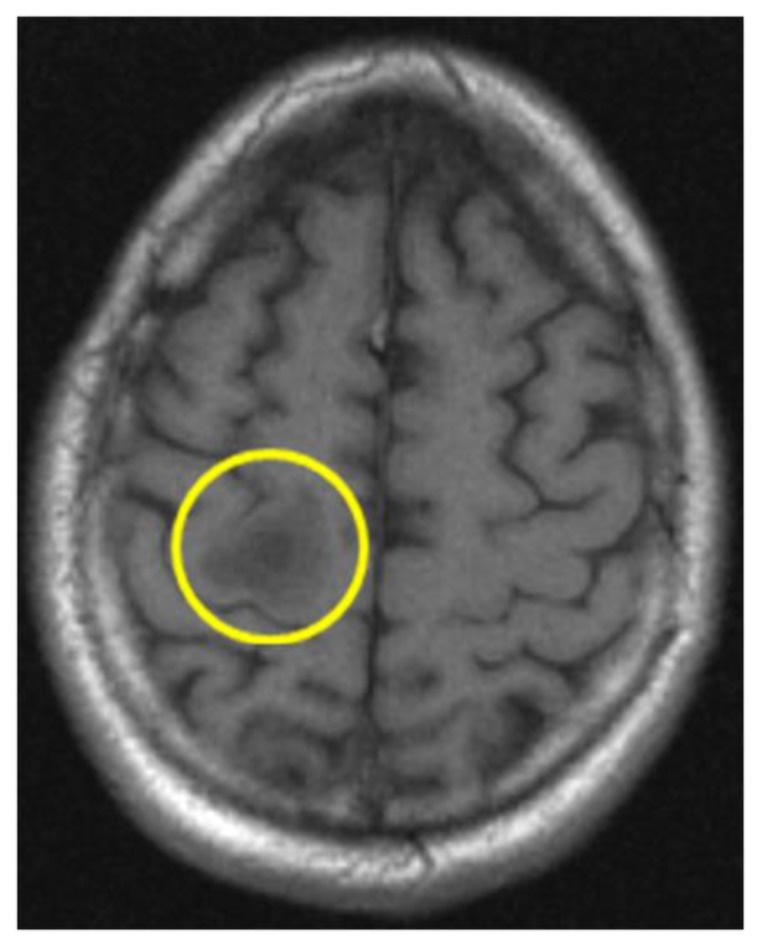
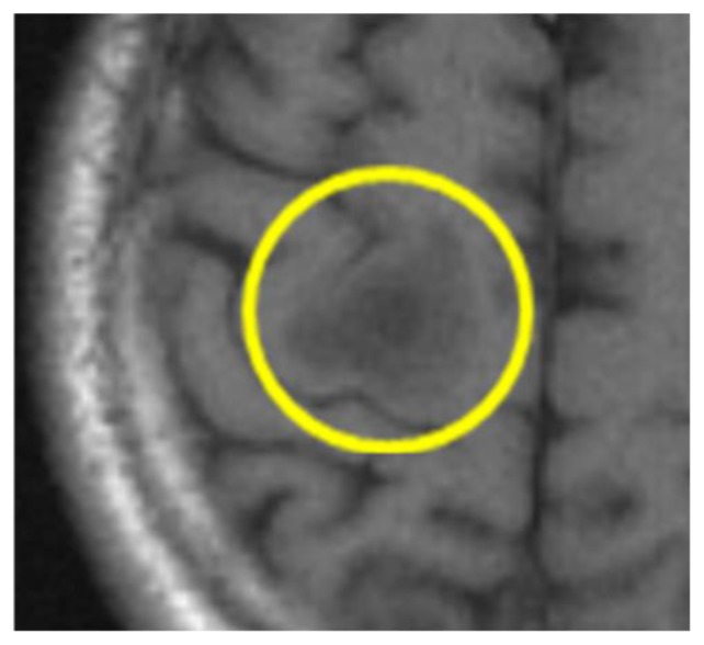
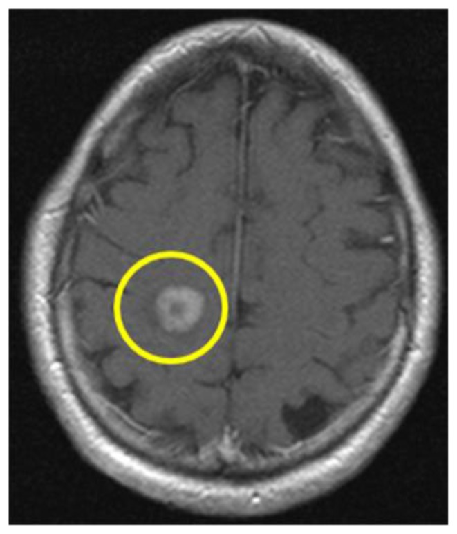
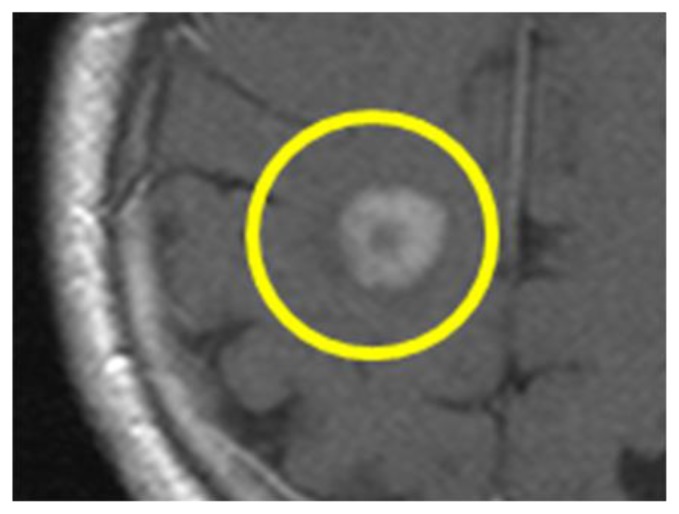
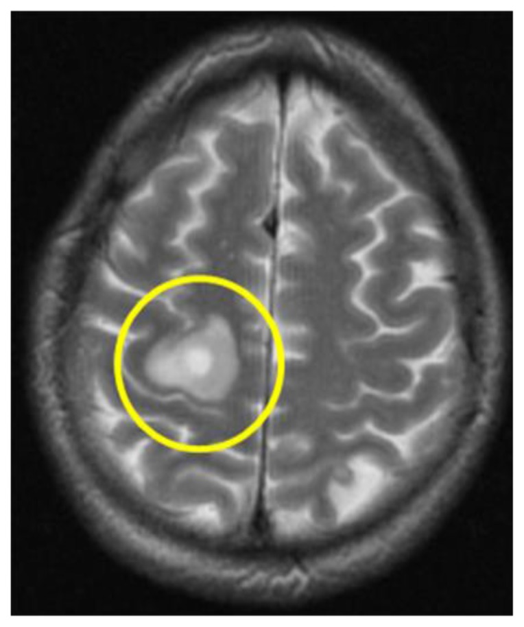
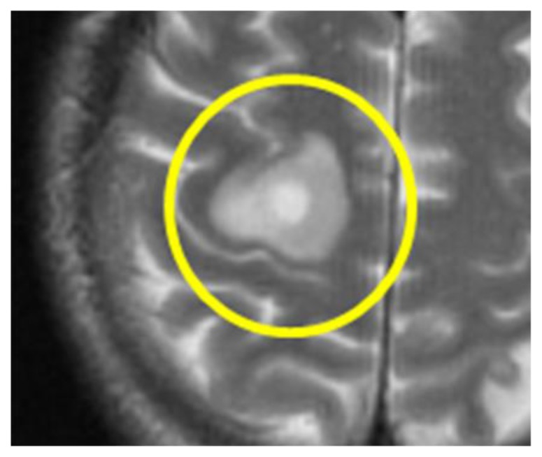
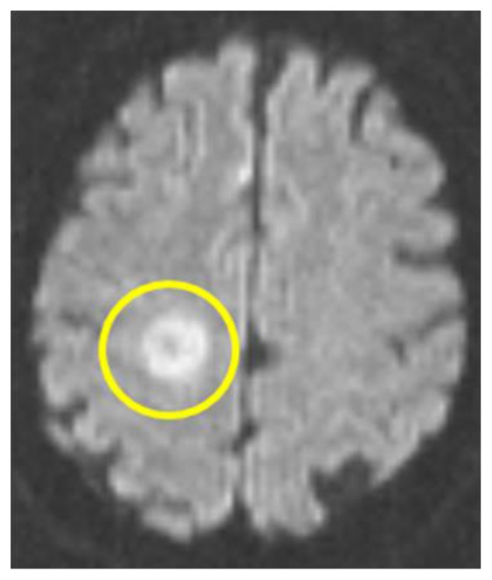
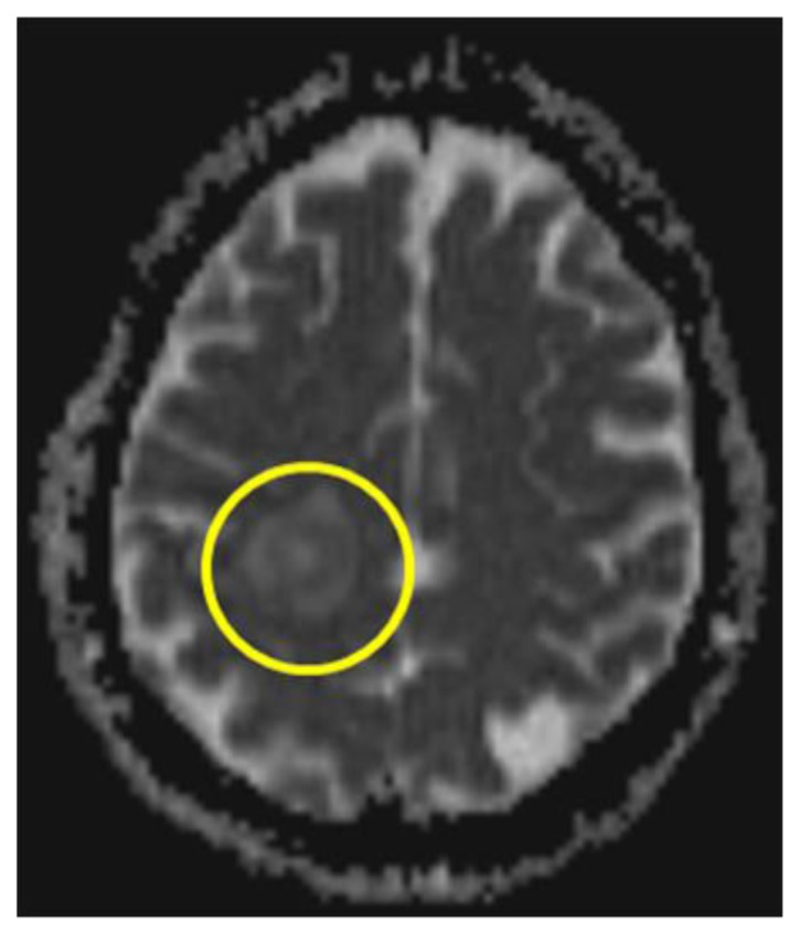
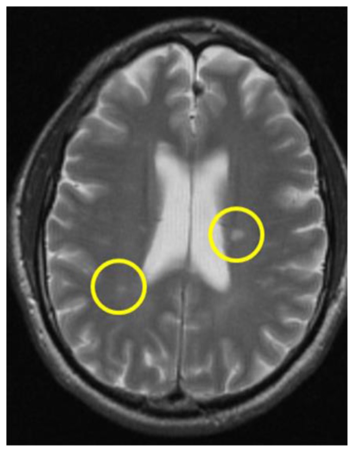
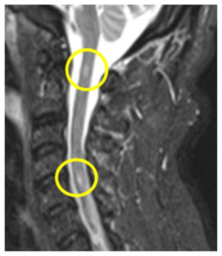
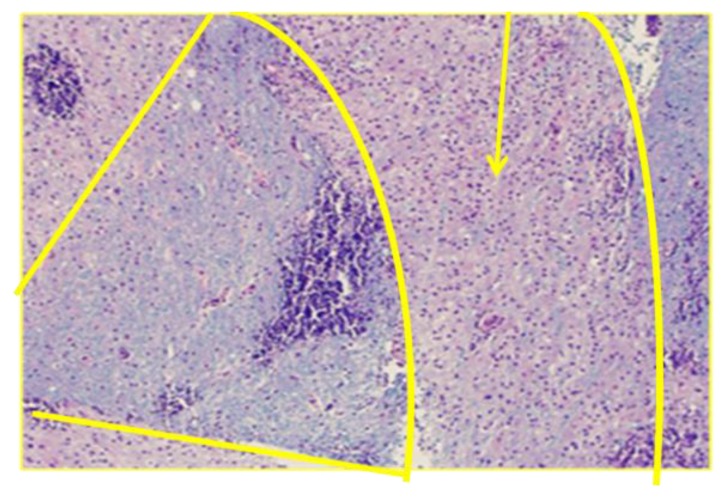
Baló's concentric sclerosis is a primary inflammatory central nervous system demyelinating disease that is considered a rare, radiographically and pathologically distinct variant of multiple sclerosis. Baló's concentric sclerosis is characterized by alternating rings of demyelinated and myelinated axons, and it is most frequently diagnosed postmortem by autopsy or, more recently, by magnetic resonance imaging without pathologic verification. This report is of a case of Baló's concentric sclerosis in which the patient presented with left-sided focal sensorimotor deficits. The patient's lesion demonstrated characteristics of Baló's concentric sclerosis by magnetic resonance imaging, but since a neoplastic process was also suspected initially, the patient underwent a surgical biopsy. This pathology sample now provides the opportunity to correlate the tissue diagnosis of demyelination with characteristic magnetic resonance imaging findings; this comparison is infrequently found in the literature.
Keywords: Baló’s concentric sclerosis; MRI; Pathology; demyelinating disease.
Figures
Similar articles
-
[Antemortem diagnosis of Balo's concentric sclerosis based on MRI. Case report].Neurol Neurochir Pol. 2002 May-Jun;36(3):565-70; discussion 570-1. Neurol Neurochir Pol. 2002. PMID: 12185812 Polish.
-
[Balò's concentric sclerosis in a North-African patient].Rev Neurol (Paris). 2005 Jan;161(1):78-80. doi: 10.1016/s0035-3787(05)84977-x. Rev Neurol (Paris). 2005. PMID: 15678005 French.
-
[Recent progress in multiple sclerosis research: astrocytopathy in demyelinating diseases].Rinsho Shinkeigaku. 2010 Nov;50(11):788-93. doi: 10.5692/clinicalneurol.50.788. Rinsho Shinkeigaku. 2010. PMID: 21921443 Review. Japanese.
-
Baló's concentric sclerosis: surviving normal myelin in a patient with a relapsing-remitting dinical course.Mult Scler. 2001 Dec;7(6):375-82. doi: 10.1177/135245850100700606. Mult Scler. 2001. PMID: 11795459
-
Astrocytopathy in Balo's disease.Mult Scler. 2011 Jul;17(7):771-9. doi: 10.1177/1352458511400475. Epub 2011 Apr 1. Mult Scler. 2011. PMID: 21459811 Review.
Cited by
-
Tumefactive Demyelinating Lesions in Multiple Sclerosis and Associated Disorders.Curr Neurol Neurosci Rep. 2016 Mar;16(3):26. doi: 10.1007/s11910-016-0626-9. Curr Neurol Neurosci Rep. 2016. PMID: 26847090 Review.
-
Baló's concentric sclerosis presenting asymptomatically in a child: clinico-radiological-pathological correlation.BMJ Case Rep. 2023 Nov 27;16(11):e256185. doi: 10.1136/bcr-2023-256185. BMJ Case Rep. 2023. PMID: 38011943 Free PMC article. No abstract available.
-
Clinical Problem-Solving: A 19-Year-Old Woman With Progressive Neurological Decline and Multiple Intracranial Lesions.Neurohospitalist. 2024 Oct;14(4):419-422. doi: 10.1177/19418744241273283. Epub 2024 Aug 13. Neurohospitalist. 2024. PMID: 39308457
-
A Rare Case of Balo Concentric Sclerosis, a Subtype of Tumefactive Multiple Sclerosis, in a 40-Year-Old Male: Case Report.Cureus. 2022 Apr 11;14(4):e24033. doi: 10.7759/cureus.24033. eCollection 2022 Apr. Cureus. 2022. PMID: 35547427 Free PMC article.
-
7 Tesla MRI of Balo's concentric sclerosis versus multiple sclerosis lesions.Ann Clin Transl Neurol. 2018 Jun 29;5(8):900-912. doi: 10.1002/acn3.572. eCollection 2018 Aug. Ann Clin Transl Neurol. 2018. PMID: 30128315 Free PMC article.
References
-
- Kavanaah EC, Heran MK, Fenton DM, Lapointe JS, Nugent RA, Graeb DA. Diffusion-weighted imaging findings in Balo concentric sclerosis. Br J Radiol. 2006 Jul;79(943):e28–31. - PubMed
-
- Li Y, Xie P, Fan X, Tang H. Baló’s concentric sclerosis presenting with benign clinical course and multiple sclerosis-like lesions on magnetic resonance images. Neurol India. 2009 Jan-Feb;57(1):66–8. - PubMed
-
- Mowry EM, Woo JH, Ances BM. Baló’s concentric sclerosis presenting as a stroke-like syndrome. Nat Clin Pract Neurol. 2007 Jun;3(6):349–54. - PubMed
Publication types
MeSH terms
LinkOut - more resources
Full Text Sources
Other Literature Sources
Medical