

doi: 10.1021/ja408513m.
Epub 2014 Jan 14.
Dynamics of soft nanomaterials captured by transmission electron microscopy in liquid water
Affiliations
- PMID: 24422495
- PMCID: PMC4021868
- DOI: 10.1021/ja408513m
Item in Clipboard
Dynamics of soft nanomaterials captured by transmission electron microscopy in liquid water
J Am Chem Soc.
.
Abstract
In this paper we present in situ transmission electron microscopy of synthetic polymeric nanoparticles with emphasis on capturing motion in a solvated, aqueous state. The nanoparticles studied were obtained from the direct polymerization of a Pt(II)-containing monomer. The resulting structures provided sufficient contrast for facile imaging in situ. We contend that this technique will quickly become essential in the characterization of analogous systems, especially where dynamics are of interest in the solvated state. We describe the preparation of the synthetic micellar nanoparticles together with their characterization and motion in liquid water with comparison to conventional electron microscopy analyses.
Figures
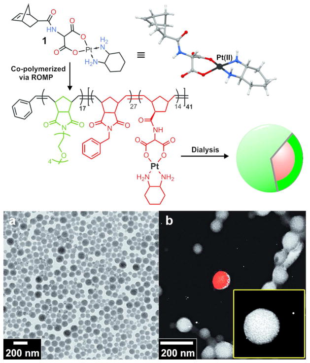
Preparation of micellar nanoparticles consisting of a Pt(II)-labeled core. TOP: Structure of Pt(II)-nobornyl monomer (1), X-ray crystal structure and amphiphilic block copolymer. Dialysis from DMF into water yielded high contrast, spherical micelles with Pt(II)-labeled cores (red). BOTTOM: a) Conventional dry-state TEM of unstained micelles. b) STEM-energy-dispersive X-ray spectroscopy (STEM-EDS) elemental map indicating platinum overlaid on the corresponding STEM-High-angle annular dark-field microscopy (HAADF) image. Inset shows zoomed STEM-HAADF image of a particle (yellow box has horizontal dimension of 200 nm).

LEFT Frames: Sequential snapshots from in situ TEM movies of nanoparticles in liquid water at times shown. Five particle centers (A–E) where selected with arrows indicating particle motion during the time lapse. RIGHT: Velocity vs. time plots for particles A and B highlighting distinct motion for each of the two connected particles. The dotted line in each graph represents the average velocity for each particle during the imaged time lapse (Average velocity, particle A = 27.5 nm/s, particle B = 18.0 nm/s). See Figure S7, Supporting Information for velocity plots for particles C, D, and E. Time displayed as minutes:seconds in movie frames. Motion and velocity analyses were performed using ImageJ with MTrackJ plugin. For movies see supporting files: Movie_Fig2_Particles_AB.mov, Movie_Fig2_Particles_CDE.mov, and Movie_S1_Fig2.avi. Images captured in silicon nitride in situ cells with 50 nm thick windows.
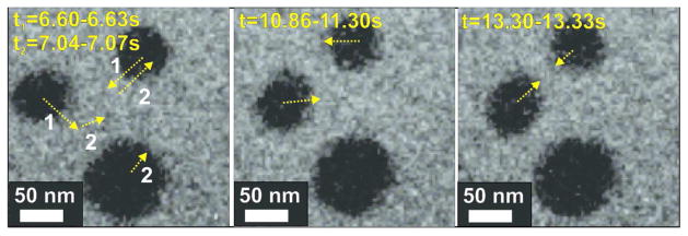
Screen shots from an in situ liquid stage movie. Arrows indicate direction of motion between the times indicated (see Movie_Fig3.mov). Time is displayed in seconds.

Comparison of Pt(II)-core micelles visualized via cryo- (left) and in situ (right) TEM showing dispersed particles for each method.
References
Publication types
MeSH terms
Substances
Grants and funding
LinkOut - more resources
Full Text Sources
Other Literature Sources
