Case Reports
. 2014 Jan 14:9:14.
doi: 10.1186/1749-8090-9-14.

A rare case of primary cardiac B cell lymphoma

Affiliations
Case Reports

A rare case of primary cardiac B cell lymphoma

Andreas Habertheuer et al. J Cardiothorac Surg. .

Abstract

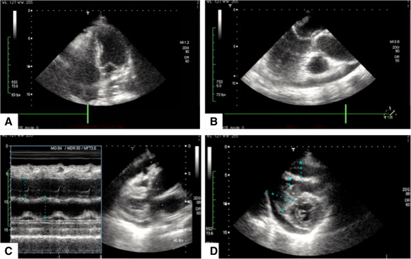
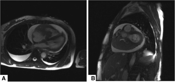
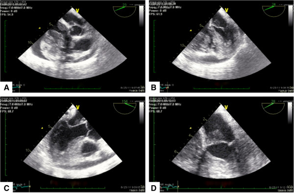
Primary cardiac lymphomas represent an extremely rare entity of extranodal lymphomas and should be distinguished from secondary cardiac involvement of disseminated lymphomas belonging to the non-Hodgkin's classification of blood cancers. Only 90 cases have been reported in literature. Presentation of cardiac lymphomas on imaging studies may not be unambiguous since they potentially mimic other cardiac neoplasms including myxomas, angiosarcoma or rhadomyomas and therefore require multimodality cardiac imaging, endomyocardial biopsy, excisional intraoperative biopsy and pericardial fluid cytological evaluation to establish final diagnosis.Herein we report the case of a 70 y/o immunocompetent Caucasian female with a rapidly progressing superior vena cava syndrome secondary to a large primary cardiac diffuse large B cell lymphoma (NHL lymphoma) almost completely obstructing the right atrium, right ventricle and affecting both mitral and tricuspid valve. The patient had no clinical evidence of disseminated disease and was successfully treated with extensive debulking during open-heart surgery on cardiopulmonary bypass and 6 cycles of rituximab, cyclophosphamide, doxorubicin, vincristine and prednisone chemotherapy (R-CHOP).


Figures

Figure 1
Figure 1
Initial transthoracic echocardiography (TTE) study showing no structural abnormalities on all views. A 4-chamber view. Regular ventricles and atria, no suspicious masses can be identified. Intact mitral and tricuspid valvular apparatus. B Parasternal long axis showing a regular, non obstructed left ventricular outflow tract. Left Atrium, mitral valve with anterior and posterior leaflet, left ventricle, aortic valve and ascending aorta. No ventricular wall dyskinesias, no obstructions. C Aortic root and left atrial diameter measurement. 35,5 mm versus 38.5 mm D Parasternal short axis. Mitral valve level showing intact valvular apparatus, no vegetations can be identified.
Figure 2
Figure 2
Magnetic resonance imaging demonstrating no evidence of mediastinal lymphadenopathy. A Axial view, T2 weighted images showing both pericardial and bilateral pleural effusions, right greater than left. Right ventricular wall irregularities and right greater than left intracavitary tumor formation, partially obstructing the right atrial and the right ventricular flow. B Sagittal view, T2 weighted images evidencing intracavitary tumor formation and both pericardial and bilateral pleural effusions. No evidence of mediastinal lymphadenopathy could be observed.
Figure 3
Figure 3
Repeat transesophageal echocardiography (TEE) study. A 5-chamber view. Tumor formation in both left and right sided cardiac structures, mitral and tricuspid valves are affected. Of note, both, the right atrium and the right ventricle are significantly obstructed. B Mid-esophageal 4-chamber view confirming extensive tumor formation and showing significant involvement of the mitral valve leaflets. Both, the right atrium and the right ventricle are severely obstructed. C Mid esophageal long axis confirming mitral valve involvement. D 4-chamber view, with cut apex. Tumor formation on mitral valve, however mitral valve apparatus does not appear to be diffusely infiltrated or misconfigured.
Figure 4
Figure 4
Histology. Complete destructive replacement of the myocardial wall by a diffuse large B cell lymphoma that demonstrated plasmacytoid differentiation and blasts.

References

    1. McAllister HA Jr, Hall RJ, Cooley DA. Tumors of the heart and pericardium. Curr Prob Cardiol. 1999;9(2):57–116. - PubMed
    1. Bisel HF, Wroblewski F, Ladue JS. Incidence and clinical manifestations of cardiac metastases. J Am Med Assoc. 1953;9(8):712–715. doi: 10.1001/jama.1953.02940250018005. - DOI - PubMed
    1. Johri A, Baetz T, Isotalo PA, Nolan RL, Sanfilippo AJ, Ropchan G. Primary cardiac diffuse large B cell lymphoma presenting with superior vena cava syndrome. Can J Cardiol. 2009;9(6):e210–e212. doi: 10.1016/S0828-282X(09)70110-2. - DOI - PMC - PubMed
    1. Miguel CE, Bestetti RB. Primary cardiac lymphoma. Int J Cardiol. 2011;9(3):358–363. doi: 10.1016/j.ijcard.2010.02.016. - DOI - PubMed
    1. Nascimento AF, Winters GL, Pinkus GS. Primary cardiac lymphoma: clinical, histologic, immunophenotypic, and genotypic features of 5 cases of a rare disorder. Am J Surg Pathol. 2007;9(9):1344–1350. doi: 10.1097/PAS.0b013e3180317341. - DOI - PubMed

Publication types

LinkOut - more resources