Review
. 2014 Jan 15;6(1):4.
doi: 10.1186/alzrt234. eCollection 2014.

Chronic traumatic encephalopathy: a spectrum of neuropathological changes following repetitive brain trauma in athletes and military personnel

Affiliations
Review

Chronic traumatic encephalopathy: a spectrum of neuropathological changes following repetitive brain trauma in athletes and military personnel

Thor D Stein et al. Alzheimers Res Ther. .

Abstract

Chronic traumatic encephalopathy (CTE) is a progressive neurodegenerative disease that occurs in association with repetitive traumatic brain injury experienced in sport and military service. In most instances, the clinical symptoms of the disease begin after a long period of latency ranging from several years to several decades. The initial symptoms are typically insidious, consisting of irritability, impulsivity, aggression, depression, short-term memory loss and heightened suicidality. The symptoms progress slowly over decades to include cognitive deficits and dementia. The pathology of CTE is characterized by the accumulation of phosphorylated tau protein in neurons and astrocytes in a pattern that is unique from other tauopathies, including Alzheimer's disease. The hyperphosphorylated tau abnormalities begin focally, as perivascular neurofibrillary tangles and neurites at the depths of the cerebral sulci, and then spread to involve superficial layers of adjacent cortex before becoming a widespread degeneration affecting medial temporal lobe structures, diencephalon and brainstem. Most instances of CTE (>85% of cases) show abnormal accumulations of phosphorylated 43 kDa TAR DNA binding protein that are partially colocalized with phosphorylated tau protein. As CTE is characterized pathologically by frontal and temporal lobe atrophy, by abnormal deposits of phosphorylated tau and by 43 kDa TAR DNA binding protein and is associated clinically with behavioral and personality changes, as well as cognitive impairments, CTE is increasingly categorized as an acquired frontotemporal lobar degeneration. Currently, some of the greatest challenges are that CTE cannot be diagnosed during life and the incidence and prevalence of the disorder remain uncertain. Furthermore, the contribution of age, gender, genetics, stress, alcohol and substance abuse to the development of CTE remains to be determined.


Figures
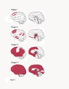
Figure 1
Figure 1
Four stages of chronic traumatic encephalopathy. Schematic of the regions involved by tau pathology in the four neuropathological stages of chronic traumatic encephalopathy.
Figure 2
Figure 2
Stage I chronic traumatic encephalopathy. In stage I chronic traumatic encephalopathy, phosphorylated tau (p-tau) pathology is found in limited discrete perivascular foci (A), typically at the depths of sulci or around small vessels (black circles). There is limited p-tau pathology in the cortex adjacent to the involved foci (B). Occasional p-tau neurites are found in the nucleus basalis of Meynert (C). There is no pathology in the amygdala (D) or CA1 of the hippocampus (E). CP-13-immunostained 50 μm tissue sections, some counterstained with cresyl violet. Scale bar = 100 μm. Adapted from [29].
Figure 3
Figure 3
Stage II chronic traumatic encephalopathy. In stage II chronic traumatic encephalopathy, there is spread of pathology from focal epicenters (A) to the superficial layers of adjacent cortex (B). The nucleus basalis of Meynert (C) shows moderate neurofibrillary tangles and neurites. The medial temporal lobe shows only mild neurofibrillary pathology, including the amygdala (D) and CA1 of the hippocampus (E). CP-13-immunostained 50 μm tissue sections, some counterstained with cresyl violet. Scale bar = 100 μm. Adapted from [29].
Figure 4
Figure 4
Stage III chronic traumatic encephalopathy. In stage III chronic traumatic encephalopathy, phosphorylated tau pathology is severe and widespread throughout the frontal, insular, temporal, and parietal cortices. The cortical epicenters and depths of the sulci often consist of confluent masses of neurofibrillary tangles (NFTs) and astrocytic tangles (A). The intervening cortices show advanced neurofibrillary degeneration (B). The nucleus basalis of Meynert shows dense NFTs (C). The amygdala (D) and hippocampus (E) show marked neurofibrillary pathology. CP-13-immunostained 50 μm tissue sections, some counterstained with cresyl violet. Scale bar = 100 μm. Adapted from [29].
Figure 5
Figure 5
Stage IV chronic traumatic encephalopathy. In stage IV chronic traumatic encephalopathy, there is widespread phosphorylated tau (p-tau) pathology affecting most regions of the cerebral cortex and medial temporal lobe with relative sparing of the calcarine cortex. Astrocytic tangles are prominent and there is marked neuronal loss in the cortex, amygdala and hippocampus. p-tau neurofibrillary tangles (NFTs) are reduced in size and density. The cortical epicenters show severe neuronal loss and prominent astrocytic tangles (A); similar changes are found throughout the frontal, temporal and parietal cortices (B). The nucleus basalis of Meynert shows marked neurofibrillary pathology and gliosis (C). The amygdala demonstrates intense gliosis and p-tau neuronal and glial degeneration (D). The hippocampus is sclerotic with marked neuronal loss, gliosis, ghost NFTs and astrocytic tangles (E). CP-13-immunostained 50 μm tissue sections, some counterstained with cresyl violet. Scale bar = 100 μm. Adapted from [29].
Figure 6
Figure 6
Chronic traumatic encephalopathy with comorbid disease. Percentage of cases with chronic traumatic encephalopathy (CTE) with and without comorbidity. Of those cases with CTE and Alzheimer’s disease, 43% had overlapping Lewy body disease; of those with CTE and frontotemporal lobar degeneration, 50% also had Lewy body disease.

References

    1. Osnato M. Postconcussion neurosis-traumatic encephalitis – a conception of postconcussion phenomena. Arch NeurPsych. 1927;6:181–214. doi: 10.1001/archneurpsyc.1927.02210020025002. - DOI
    1. Martland H. Punch drunk. JAMA. 1928;6:1103–1107. doi: 10.1001/jama.1928.02700150029009. - DOI
    1. Millsbaugh JA. Dementia pugilistica. US Naval Med Bulletin. 1937;6:297–361.
    1. Critchley M. Punch-Drunk Syndromes: the Chronic Traumatic Encephalopathy of Boxers. Hommage a Clovis Vincent. Paris: Maloin; 1949.
    1. McCown IA. Protecting the boxer. JAMA. 1959;6:1409–1413. doi: 10.1001/jama.1959.03000300005002. - DOI - PubMed