. 2014 Jan 15;16(1):R10.
doi: 10.1186/ar4436.

Intra-articular injection of collagenase in the knee of rats as an alternative model to study nociception associated with osteoarthritis

Intra-articular injection of collagenase in the knee of rats as an alternative model to study nociception associated with osteoarthritis

Sara Adães et al. Arthritis Res Ther. .

Abstract

Introduction: Animal models currently used in osteoarthritis-associated pain research inadequately reproduce the initiating events and structural pathology of human osteoarthritis. Conversely, intra-articular injection of collagenase is a structurally relevant model, as it induces articular degeneration both by digesting collagen from cartilage and by causing articular instability, thereby reproducing some of the main events associated with osteoarthritis onset and development. Here, we evaluated if the intra-articular injection of collagenase can be an alternative model to study nociception associated with osteoarthritis.

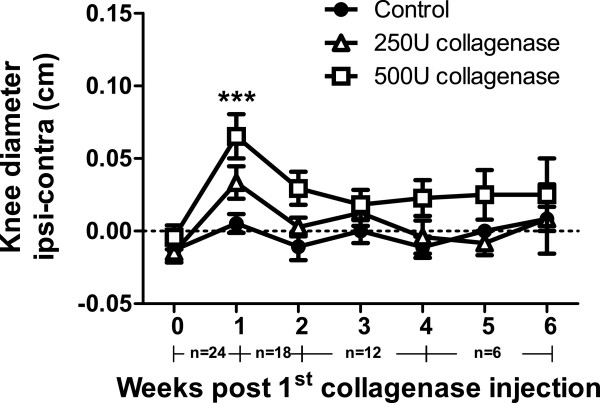
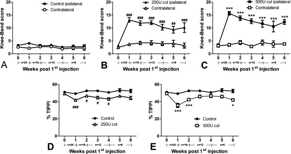
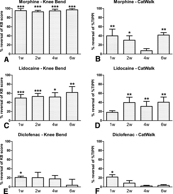
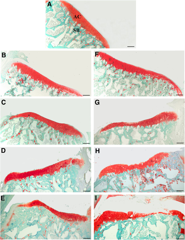
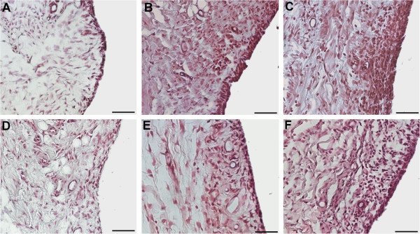
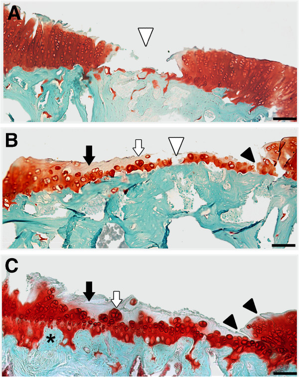
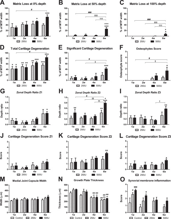
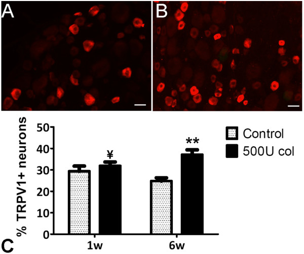
Methods: Osteoarthritis was induced by two intra-articular injections of either 250 U or 500 U of collagenase into the left knee joint of adult male Wistar rats. A six weeks time-course assessment of movement- and loading-induced nociception was performed by the Knee-Bend and CatWalk tests. The effect of morphine, lidocaine and diclofenac on nociceptive behaviour was evaluated in animals injected with 500 U of collagenase. Joint histopathology was scored for both doses throughout time. The expression of transient receptor potential vanilloid 1 (TRPV1) in ipsilateral dorsal root ganglia (DRG) was evaluated.

Results: An increase in nociceptive behaviour associated with movement and loading of affected joints was observed after intra-articular collagenase injection. With the 500 U dose of collagenase, there was a significant correlation between the behavioural and the histopathological osteoarthritis-like structural changes developed after six weeks. One week after injection of 500 U collagenase, swelling of the injected knee and inflammation of the synovial membrane were also observed, indicating the occurrence of an early inflammatory reaction. Behavioural changes induced by the 500 U dose of collagenase were overall effectively reversed by morphine and lidocaine. Diclofenac was effective one week after injection. TRPV1 expression increased six weeks after 500 U collagenase injection.

Conclusion: We conclude that the intra-articular injection of 500 U collagenase in the knee of rats can be an alternative model for the study of nociception associated with osteoarthritis, since it induces significant nociceptive alterations associated with relevant osteoarthritis-like joint structural changes.


Figures

Figure 1
Figure 1
Inflammation index. Joint swelling assessed by measuring knee diameters (difference between ipsilateral and contralateral knees). Collagenase-injected animals present increased knee diameters one week after injection. The number of animals per group at each time-point is indicated below the graphs. An initial number of 24 animals was used in each group. Animals were then sacrificed at different time-points for histopathology (one, two, four and six weeks; n = 6 per dose and per time-point). Mean ± SEM, two-way ANOVA followed by Bonferroni post-hoc test for comparisons between groups at each time-point. ***P <0.001, for comparisons between control and the 500 U collagenase group.
Figure 2
Figure 2
Nociceptive behaviour. Nociception induced by movement and loading in control and collagenase-injected rats evaluated by the Knee-Bend and CatWalk tests. Knee-Bend score (A-C) is presented for both ipsilateral and contralateral knees of saline-injected control rats (A), and rats injected with 250 U (B) or 500 U (C) of collagenase. CatWalk data (D, E) are expressed as the percentage of total ipsilateral paw print intensity (%TIPPI). Collagenase-injected animals present increased movement- and loading-induced nociception. The number of animals per group at each time-point is indicated below the graphs. An initial number of 24 animals was used in each group. Animals were then sacrificed at different time-points for histopathology (one, two, four and six weeks; n = 6 per dose and per time-point). Mean ± SEM, two-way ANOVA followed by Bonferroni post-hoc test for comparisons between groups at each time-point. #P <0.05, ###P <0.001, for comparisons between control and the 250 U collagenase group; *P <0.05, ***P <0.001, for comparisons between control and the 500 U collagenase group.
Figure 3
Figure 3
Pharmacological evaluation. Effect of morphine (6 mg/Kg, s.c., A, B), lidocaine (5 mg, i.a., C, D) or diclofenac (30 mg/Kg, p.o., E, F) on movement- and loading-induced nociception assessed by the Knee-Bend (A,C,E) and the CatWalk (B,D,F) tests, at one, two, four and six weeks after injection of 500 U of collagenase. A: Morphine effectively reversed the Knee-Bend scores at all time-points of disease progression. B: Morphine was effective at one, two and six weeks of disease progression. C: Lidocaine effectively reversed the Knee-Bend scores at all time-points of disease progression. D: Lidocaine was effective at two, four and six weeks of disease progression. E: Diclofenac effectively reversed the Knee-Bend scores one week after injection. F: Diclofenac was effective one week after injection. Mean ± SEM. Repeated measures ANOVA followed by Dunnett’s post-hoc test; *P <0.05, **P <0.01, ***P <0.001.
Figure 4
Figure 4
Progression of histopathological changes in knee joint sections stained with Fast Green and Safranin O.A: Control animals show unaltered articular cartilage (AC) and subchondral bone (SB). Animals injected with 250 U collagenase (B, C, D, E) show increased thinning of the articular cartilage throughout time, along with focal chondrocyte disorganization. Animals injected with 500 U (F, G, H, I) show signs of OA onset at week 4 (H) that further develop until week 6 (I) when extensive damage of the articular cartilage is observed. Bar: 50 μm.
Figure 5
Figure 5
Synovial membrane histopathology. Images representative of the synovial membrane inflammation one week (A-C) and six weeks (D-F) after injection of saline (A,D), 250 U (B, E) or 500 U of collagenase (C, F). An increased number of synovial lining cell layers, proliferation of subsynovial tissue and infiltration of inflammatory cells are observed. Changes were most pronounced with the 500 U dose, one week after injection, receding thereafter. Bar: 50 μm.
Figure 6
Figure 6
Articular cartilage degeneration six weeks after the injection of 500 U of collagenase. Extensive cartilage degeneration, comprising areas of marked erosion with exposure of the subchondral bone (white arrowhead; A, B), loss of proteoglycan staining and cell depletion (black arrow; B, C), chondrocyte clustering and hypertrophy (white arrow; B, C), fissures (black arrowhead; B, C) and tidemark undulation (*; C). Bar: 50 μm.
Figure 7
Figure 7
Histopathological scoring. Histopathological assessment of cartilage degenerative changes in the medial tibial plateau (MTP) of control animals and animals injected with 250 or 500 U collagenase, one, two, four and six weeks after injection. The following parameters were evaluated: cartilage matrix loss width (CMLW) measured along the surface (0% depth, A), the midzone (50% depth, B) and tidemark (100% depth, C); total cartilage degeneration width (TCDW, D); significant cartilage degeneration width (SCDW, E); osteophyte score (OS, F); zonal depth ratio (ZDR) of lesions at zone 1 (at the medial edge of the joint, G), zone 2 (at the centre of the MTP, H) and zone 3 (adjacent to the cruciate ligaments, I); cartilage degeneration score (CDS) at zones 1 (J), 2 (K) and 3 (L); synovial membrane inflammation score (SMIS, M); medial joint capsule width (MJCW, N); growth plate thickness (GPT, O). Mean ± SEM. Measured parameters were analysed by one-way ANOVA followed by Tukey post-hoc test; scored parameters were analysed by the Kruskal-Wallis test with Dunn’s post-hoc test. *P <0.05, **P <0.01, ***P <0.001, for comparisons between collagenase injected and control animals within each time point; +P <0.05, +++P <0.001, for comparisons between the 250 and 500 U groups within each time-point; #P <0.05, ###P <0.001, for comparisons between week 1 and week 6 within each group; P <0.05, ‡‡P <0.01, ‡‡‡P <0.001, for comparisons between week 2 and week 6 within each group; P <0.05, ††P <0.01, †††P <0.001, for comparisons between week 4 and week 6 within each group; ¥P <0.05, ¥¥P <0.01, ¥¥¥P <0.001, between collagenase injected and control animals from a different time point.
Figure 8
Figure 8
TRPV1 expression. Transient receptor potential vanilloid 1 (TRPV1) expression was evaluated in the L3, L4 and L5 ipsilateral dorsal root ganglia (DRG) of control and 500 U collagenase injected rats. (A, B) TRPV1 expressed in small-sized L4 DRG neurons of control rats (A) and 500 U collagenase-injected rats (B), six weeks after injection. C: Six weeks after the injection of 500 U of collagenase there is an increase in the expression of TRPV1. Mean ± SEM. **P <0.01, significantly different from control rats at the same time-point. ¥P <0.05, significantly different from control rats at a different time-point.

References

    1. WHO Scientific Group on the Burden of Musculoskeletal Conditions at the Start of the New Millennium. The burden of musculoskeletal conditions at the start of the new millennium. World Health Organ Tech Rep Ser. 2003;16:i–x. 1–218, back cover. - PubMed
    1. Creamer P, Lethbridge-Cejku M, Hochberg MC. Where does it hurt? Pain localization in osteoarthritis of the knee. Osteoarthritis Cartilage. 1998;16:318–323. - PubMed
    1. Ameye LG, Young MF. Animal models of osteoarthritis: lessons learned while seeking the “Holy Grail”. Curr Opin Rheumatol. 2006;16:537–547. - PubMed
    1. Bendele A. Animal models of osteoarthritis. J Musculoskelet Neuronal Interact. 2001;16:363–376. - PubMed
    1. Bendele A. Animal models of osteoarthritis in an era of molecular biology. J Musculoskelet Neuronal Interact. 2002;16:501–503. - PubMed

Publication types