

Hippocampal sclerosis in dementia, epilepsy, and ischemic injury: differential vulnerability of hippocampal subfields
- PMID: 24423638
- PMCID: PMC4076969
- DOI: 10.1097/OPX.0000000000000170
Hippocampal sclerosis in dementia, epilepsy, and ischemic injury: differential vulnerability of hippocampal subfields
Abstract
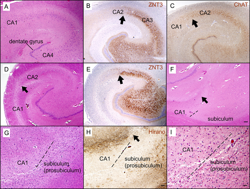
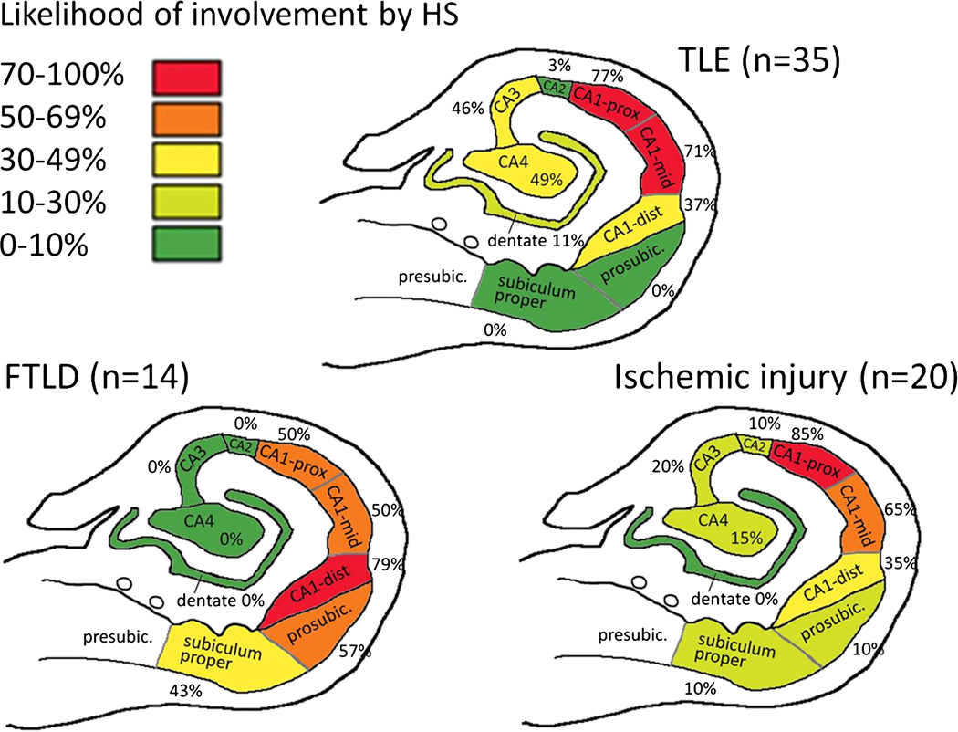
Severe neuronal loss in the hippocampus, that is, hippocampal sclerosis (HS), can be seen in 3 main clinical contexts: dementia (particularly frontotemporal lobar degeneration [FTLD]), temporal lobe epilepsy (TLE), and hippocampal ischemic injury (H-I). It has been suggested that shared pathogenetic mechanisms may underlie selective vulnerability of the hippocampal subfields such as the CA1 in these conditions. We determined the extent of neuronal loss in cases of HS-FTLD (n=14), HS-TLE (n=35), and H-I (n=20). Immunohistochemistry for zinc transporter 3 was used to help define the CA3/CA2 border in the routinely processed human autopsy tissue samples. The subiculum was involved in 57% of HS-FTLD, 10% of H-I, and 0% of HS-TLE cases (p<0.0001). The CA regions other than CA1 were involved in 57% of HS-TLE, 30% of H-I, and 0% of HS-FTLD cases (p=0.0003). The distal third of CA1 was involved in 79% of HS-FTLD, 35% of H-I, and 37% of HS-TLE cases (p=0.02). The distal third of CA1 was the only area involved in 29% of HS-FTLD, 3% of HS-TLE, and 0% of H-I cases (p=0.019). The proximal-middle CA1 was the only area affected in 50% of H-I, 29% of HS-TLE, and 0% of HS-FTLD cases (p=0.004). These findings support heterogeneity in the pathogenesis of HS.
Figures
Similar articles
-
Distinct increased metabotropic glutamate receptor type 5 (mGluR5) in temporal lobe epilepsy with and without hippocampal sclerosis.Hippocampus. 2013 Dec;23(12):1212-30. doi: 10.1002/hipo.22160. Epub 2013 Jul 23. Hippocampus. 2013. PMID: 23804486 Free PMC article.
-
Drebrin expression patterns in patients with refractory temporal lobe epilepsy and hippocampal sclerosis.Epilepsia. 2020 Aug;61(8):1581-1594. doi: 10.1111/epi.16595. Epub 2020 Jul 14. Epilepsia. 2020. PMID: 32662890
-
Manual Hippocampal Subfield Segmentation Using High-Field MRI: Impact of Different Subfields in Hippocampal Volume Loss of Temporal Lobe Epilepsy Patients.Front Neurol. 2018 Nov 20;9:927. doi: 10.3389/fneur.2018.00927. eCollection 2018. Front Neurol. 2018. PMID: 30524352 Free PMC article.
-
Hippocampal sclerosis: A review on current research status and its mechanisms.Ageing Res Rev. 2025 Jun;108:102716. doi: 10.1016/j.arr.2025.102716. Epub 2025 Mar 8. Ageing Res Rev. 2025. PMID: 40058463 Review.
-
Surgical pathology of epilepsy-associated non-neoplastic cerebral lesions: a brief introduction with special reference to hippocampal sclerosis and focal cortical dysplasia.Neuropathology. 2013 Aug;33(4):442-58. doi: 10.1111/neup.12028. Epub 2013 Mar 27. Neuropathology. 2013. PMID: 23530853 Free PMC article. Review.
Cited by
-
FTLD-TDP With and Without GRN Mutations Cause Different Patterns of CA1 Pathology.J Neuropathol Exp Neurol. 2019 Sep 1;78(9):844-853. doi: 10.1093/jnen/nlz059. J Neuropathol Exp Neurol. 2019. PMID: 31361008 Free PMC article.
-
Limbic Predominant Age-Related TDP-43 Encephalopathy (LATE): Clinical and Neuropathological Associations.J Neuropathol Exp Neurol. 2020 Mar 1;79(3):305-313. doi: 10.1093/jnen/nlz126. J Neuropathol Exp Neurol. 2020. PMID: 31845964 Free PMC article.
-
Proteomic differences in the hippocampus and cortex of epilepsy brain tissue.Brain Commun. 2021 Mar 9;3(2):fcab021. doi: 10.1093/braincomms/fcab021. eCollection 2021. Brain Commun. 2021. PMID: 34159317 Free PMC article.
-
Global Cognitive Impairment in Systemic Lupus Erythematosus Patients: A Structural MRI Study.Clin Neuroradiol. 2017 Mar;27(1):23-29. doi: 10.1007/s00062-015-0397-8. Epub 2015 May 13. Clin Neuroradiol. 2017. PMID: 25967601
-
The hippocampus: detailed assessment of normative two-dimensional measurements, signal intensity, and subfield conspicuity on routine 3T T2-weighted sequences.Surg Radiol Anat. 2017 Oct;39(10):1149-1159. doi: 10.1007/s00276-017-1843-x. Epub 2017 Mar 12. Surg Radiol Anat. 2017. PMID: 28286911
References
-
- Kim JH. Pathology of epilepsy. Exp Mol Pathol. 2001;70:345–367. - PubMed
-
- Thom M, Liagkouras I, Elliot KJ, et al. Reliability of patterns of hippocampal sclerosis as predictors of postsurgical outcome. Epilepsia. 2010;51:1801–1808. - PubMed
-
- Leifer D, Kowall NW. Immunohistochemical patterns of selective cellular vulnerability in human cerebral ischemia. J Neurol Sci. 1993;119:217–228. - PubMed
Publication types
MeSH terms
Grants and funding
LinkOut - more resources
Full Text Sources
Other Literature Sources
Medical
Miscellaneous
