. 2014 Apr;42(6):3590-606.
doi: 10.1093/nar/gkt1379. Epub 2014 Jan 13.

AUF1 contributes to Cryptochrome1 mRNA degradation and rhythmic translation

Affiliations

AUF1 contributes to Cryptochrome1 mRNA degradation and rhythmic translation

Kyung-Ha Lee et al. Nucleic Acids Res. 2014 Apr.

Abstract

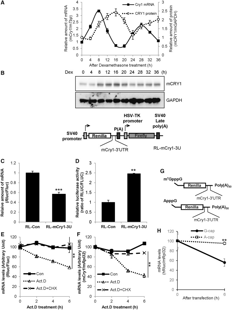
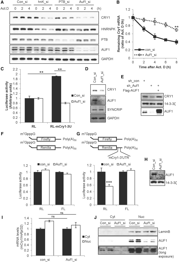
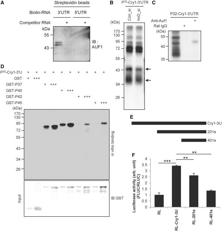
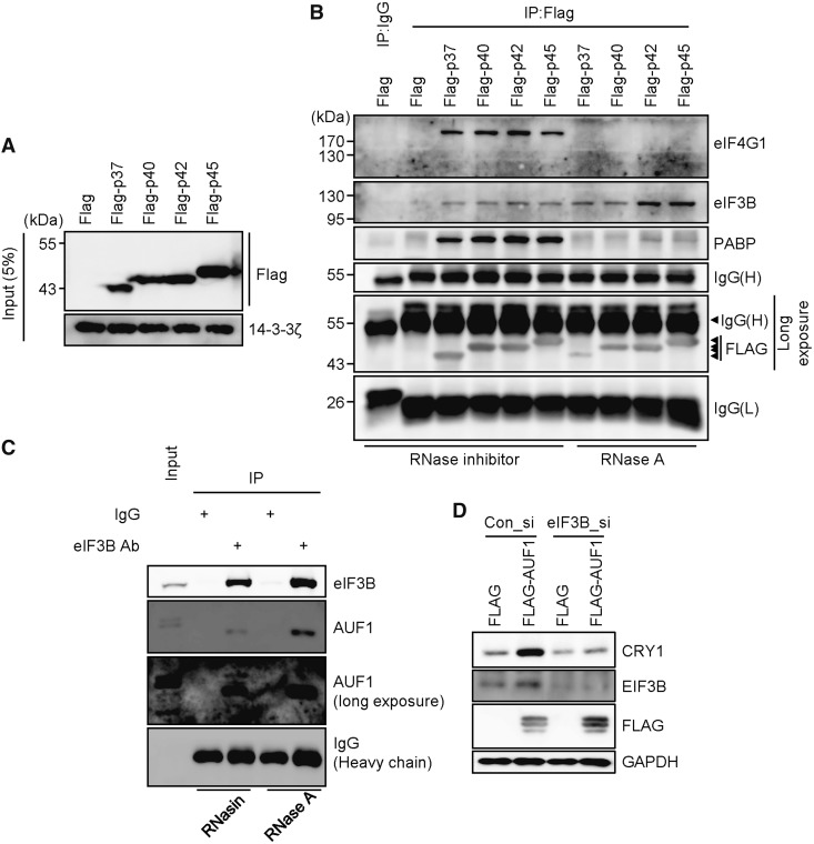
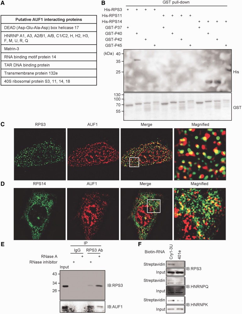
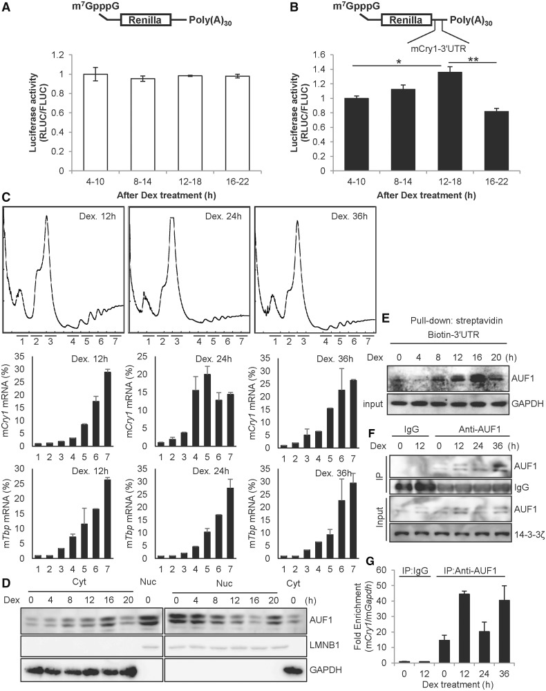
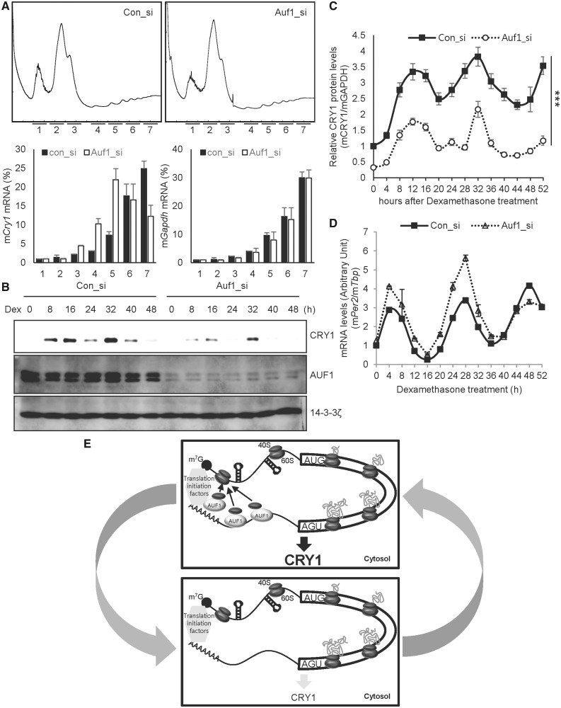
In the present study, we investigated the 3' untranslated region (UTR) of the mouse core clock gene cryptochrome 1 (Cry1) at the post-transcriptional level, particularly its translational regulation. Interestingly, the 3'UTR of Cry1 mRNA decreased its mRNA levels but increased protein amounts. The 3'UTR is widely known to function as a cis-acting element of mRNA degradation. The 3'UTR also provides a binding site for microRNA and mainly suppresses translation of target mRNAs. We found that AU-rich element RNA binding protein 1 (AUF1) directly binds to the Cry1 3'UTR and regulates translation of Cry1 mRNA. AUF1 interacted with eukaryotic translation initiation factor 3 subunit B and also directly associated with ribosomal protein S3 or ribosomal protein S14, resulting in translation of Cry1 mRNA in a 3'UTR-dependent manner. Expression of cytoplasmic AUF1 and binding of AUF1 to the Cry1 3'UTR were parallel to the circadian CRY1 protein profile. Our results suggest that the 3'UTR of Cry1 is important for its rhythmic translation, and AUF1 bound to the 3'UTR facilitates interaction with the 5' end of mRNA by interacting with translation initiation factors and recruiting the 40S ribosomal subunit to initiate translation of Cry1 mRNA.


Figures

Figure 1.
Figure 1.
The 3′UTR of Cry1 is involved in translation. (A) NIH 3T3 cells were treated with dexamethasone (Dex), and cells were subjected to mRNA quantification or immunoblotting at the indicated time points. The relative Cry1 mRNA levels were expressed as the mean ± SEM (closed squares/solid line). The relative mCRY1 protein level (open circles/dotted line) were normalized to GAPDH and plotted. mCry1 mRNA (CircWave, P = 1 × 10⁻⁷) and mCRY1 protein (CircWave, P = 7 × 10⁻⁷) levels between 8–36h are significantly rhythmic. (B) Cry1-3′UTR was fused to Renilla luciferase (RL-Cry1-3U). Firefly luciferase was used as an internal control. (C) RL-con, which lacks the 3′UTR sequence, or RL-Cry1-3U plasmids were transfected into NIH 3T3 cells. After a 24-h incubation, total RNA was prepared, and mRNA levels were quantified by real-time PCR with Rluc- or Fluc-specific primers. mRNA levels were normalized to Fluc mRNA levels. The relative mRNA level of RL-con was set to 1 (n = 4, ***P < 0.0001). (D) From the same extracts of panel C, the luciferase assay was performed. Renilla luciferase activity (RLUC) was normalized to Firefly luciferase activity (FLUC), and RL-con activity (ratio of RLUC/FLUC) was set to 1 (n = 3, **P = 0.0059). (E) The Cry1-3′UTR reporter (RL-Cry1-3U) was transfected into NIH 3T3 cells. After a 24-h incubation, cells were treated with dimethyl sulfoxide (DMSO) vehicle (Con), actinomycin D (Act. D) or Act. D plus cycloheximide (CHX), and harvested at indicated time points. mRNA levels of Rluc or Fluc were measured by real-time PCR with Rluc- or Fluc-specific primers. (n = 3, **P < 0.05) (F) Untransfected NIH 3T3 cells were treated with DMSO, Act.D or Act.D plus CHX as shown in panel E. Endogenous Cry1 mRNA levels were determined by real-time PCR and normalized to Rpl32 mRNA levels at the indicated time points. The initial relative level of Cry1 mRNA was arbitrarily set to 100 (n = 3, **P < 0.05). (G) Schematic diagrams of the mRNA reporter of Cry1-3′UTR shows m7GpppG, ApppG and the 30-nt-long poly(A) tail [poly(A)30]. (H) m7GpppG and ApppG reporter mRNAs were transiently transfected and incubated 6 h, then mRNA levels were quantified. The initial relative mRNA levels of ApppG were set to 100 (n = 3, **P = 0.0021).
Figure 2.
Figure 2.
AUF1 regulates translation of Cry1. (A) Control siRNA (Con_si) or gene-specific siRNAs for HnrnpK (hnK_si), Ptbp1 (PTB_si) or Auf1 (Auf1_si) were transfected into NIH 3T3 cells. After a 12-h incubation, cells were treated with Act.D, and were harvested at the indicated time points and subjected to immunoblotting. (B) By using the same samples as those used in panel A, mRNA levels were quantified by real-time PCR with Cry1- or Rpl32-specific primers. Cry1 levels were normalized to levels of Rpl32, and the zero time point was set to 1 (n = 3, **P = 0.0033). (C) RL or RL-Cry1-3U reporter plasmids were co-transfected with control siRNA or Auf1_si into NIH 3T3 cells, and were subjected to the luciferase assay. The value of RL with con_si was set to 1 (n = 3, **P < 0.001). (D) Knock-down of Auf1 shown in panel C was confirmed by immunoblotting with indicated antibodies. (E) Plasmids that express control (sh_con) or Auf1 targeting (sh_Auf1) shRNA were transfected with Flag-tagged all AUF1 isoforms into NIH 3T3 cells. After 24-h incubation, cells were subjected to immunoblotting. (F and G) Schematic diagram of the mRNA reporter of Cry1-3′UTR shows 7-methyl-guanosine (m7G) and the 30-nt-long poly(A) tail [poly(A)30]. Firefly mRNA reporters for normalization were also used. Con_si or Auf1_si was transfected into NIH 3T3 cells and incubated for 12-h. Subsequently, cells were transfected with mRNA reporters harbouring Cry1-3′UTR or no UTR. After a 6-h incubation, cells were subjected to the luciferase assay (n = 3, P = 0.0001). (H) Immunoblotting was performed with specific antibodies using cell extracts used in panel F or G. (I) NIH 3T3 cells transfected with Con_si or Auf1_si was incubated for 12 h, and were fractionated into cytoplasmic and nuclear parts. mRNA levels were quantified by real-time PCR using Cry1- or Rpl32-specific primers. (J) Immunoblotting was performed with samples used in panel I. Lamin B expression was checked to verify fractionation.
Figure 3.
Figure 3.
AUF1 specifically binds to Cry1-3′UTR. (A) The in vitro transcribed Cry1 3′UTR or 5′UTR constructs were labelled with biotin-UTP and incubated with NIH 3T3 cell cytoplasmic extract. Streptavidin-affinity purified samples were separated by SDS-PAGE and subjected to immunoblotting with anti-AUF1. Abundant AUF1 was detected in the reaction with biotin-labelled mRNA. AUF1 binding decreased in the presence of 5-fold excess of non-labelled 3′UTR mRNA. (B) Radiolabelled 3′UTR of Cry1 was transcribed in vitro and subjected to in vitro binding and UV-crosslinking with nuclear extracts of Con_si- or Auf1_si-transfected NIH 3T3 cells. Samples were separated by SDS-PAGE for autoradiography. (C) Cytoplasmic extracts labelled by UV cross-linking with radiolabelled 3′UTR of mCry1 were subjected to immunoprecipitation with AUF1-specific antibody or normal Rat IgG as a control and then separated by SDS-PAGE for autoradiography. (D) In vitro transcribed 3′UTR was subjected to in vitro binding and UV-crosslinking assay with purified GST-tagged AUF1 isoforms (GST-P37, GST-P40, GST-P42 and GST-P45), and autoradiographic intensities were checked. In the lower panel, input levels were checked by immunoblotting with anti-GST. (E) Schematic diagram of the serially deleted mutation strategy. (F) Each deletion construct derived from full-length Cry1-3′UTR was transfected into NIH 3T3 cells, and luciferase assays were performed. The graph shows the relative luciferase activity derived from the RLUC/FLUC ratio (n = 3, ***P < 0.0001, **P < 0.001).
Figure 4.
Figure 4.
AUF1 interacts with translation initiation factors. (A) NIH 3T3 cells were transiently transfected with Flag-tagged AUF1 isoforms Flag-P37, Flag-P40, Flag-P42 and Flag-P45. After a 24-h incubation, cells were subjected to immunoblotting. (B) With the same samples used in panel A, immunoprecipitation was performed with a Flag-specific antibody under RNA-containing or RNA-free conditions, and samples were subjected to immunoblotting. (C) Untransfected NIH 3T3 cells were subjected to immunoprecipitation with anti-EIF3B after RNase inhibitor or RNase A treatment. Immunoblotting was performed with specific antibodies. (D) Con_si or eIF3B targeting siRNAs (eIF3B_si) were transfected with Flag or all Flag-AUF1 isoforms into NIH 3T3 cells. After 24-h incubation, cells were subjected to immunoblotting with annotated antibodies.
Figure 5.
Figure 5.
AUF1 directly interacts with ribosomal proteins. (A) The table for putative AUF1 interacting proteins was made base on the LTQ-orbitrap data. The full LTQ-orbitrap data file is in the Supplemental material. (B) Recombinant proteins of GST-AUF1 isoforms, His-RPS3, His-RPS11 or His-RPS14 were incubated and pulled down using GST-binding resins. Two micrograms of Histidine tag-fused RPS proteins or GST-fused AUF1 isoforms were applied. Each protein separated by SDS-PAGE was detected by the indicated antibodies. (C and D) Immunostaining was performed with AUF1-, RPS3-, or RPS14-specific antibodies. RPS3 or RPS14 was visualized using Alexa 488-conjugated secondary antibody. For AUF1, Alexa 594-conjugated secondary antibody was used for visualization with a super-resolution illumination microscopy (SIM). (E) NIH 3T3 cells were subjected to immunoprecipitation under RNA-free or RNA-containing conditions with RPS3-specific antibody, and samples were subjected to immunoblotting with indicated antibodies. (F) The in vitro-transcribed full-length or 401e with a truncated Cry1-3′UTR were labelled with biotin-UTP and incubated with NIH 3T3 cell cytoplasmic extract. Streptavidin-affinity purified samples were subjected to immunoblotting with indicated antibodies.
Figure 6.
Figure 6.
Rhythmic cytoplasmic AUF1 regulates time-dependent Cry1 translation. (A) NIH 3T3 cells were treated with dexamethasone (Dex), and the mRNA reporters lacking 3′UTR sequences were transiently transfected for 6 h at the indicated times, followed by measurement of luciferase activity. The relative values at 4–10 h were set to 1. (B) The mRNA reporters harbouring Cry1-3′UTR were transfected into Dex-treated NIH 3T3 cells (n = 4, P = 0.0097). (C) NIH 3T3 cells were treated with dexamethasone. After 12, 24, or 36 h of incubation, the cells were treated with cycloheximide. Then, the ribosomal distributions in sucrose density gradients were analysed in cell extracts (upper row). RNA samples were purified from fractions in the sucrose gradient. The amounts of mCry1 mRNA (middle row) and Tbp mRNA (bottom row) across the gradient were analysed by real-time PCR, and the relative amounts of RNA in each fraction are depicted by corresponding bars in the graphs. (D) NIH 3T3 cells were treated with 100 nM Dex and harvested at the indicated times, and cytoplasmic or nuclear extract was prepared. Next, immunoblotting was performed with specific antibodies. (E) NIH 3T3 cells were treated with Dex and harvested at the indicated times, and cytoplasmic extracts were prepared. Dex-treated cytoplasmic extracts were incubated with biotin-labelled Cry1-3′UTR, and samples were subjected to immunoblotting. (F) NIH 3T3 cells were treated with dexamethasone, and cytosolic extracts were prepared. Immunoprecipitation was performed using anti-AUF1 antibody and normal Rat IgG as a control. (G) The co-immunoprecipitated mRNAs with AUF1 shown in panel F were analysed by real-time PCR. AUF1-bound Cry1 mRNA levels were normalized to the levels of background Gapdh mRNA. The relative Cry1 mRNA level that immunoprecipitated with IgG at 0-h was set to 1.
Figure 7.
Figure 7.
AUF1 regulates circadian expression of CRY1. (A) NIH cells were transfected with control (Con_si) or Auf1-specific siRNA (Auf1_si). After 24 h of incubation, the cells were treated with cycloheximide. Then, the ribosomal distribution in sucrose gradients were analysed in cell extracts (first row). Distribution of mRNA in sucrose gradients were analysed by real-time PCR, and the amounts of RNA in each fraction are depicted by corresponding bars in the graphs. (B) NIH 3T3 cells transfected with siRNAs targeting Auf1 (Auf1_si) or control siRNA (con_si) by microporation were incubated for 12 h and harvested at the indicated time points after treatment with Dex. Immunoblotting was performed with the indicated antibodies. (C) The relative mCRY1 levels in con_si transfected (closed squares/solid line) and Auf1_si transfected (open circles/dotted line) NIH 3T3 cells. mCRY1 protein levels were normalized to GAPDH and plotted. The P-value of variance between con_si and Auf1_si group was 0.0001. mCRY1 protein levels were significantly different between con_si and Auf1_si transfected groups at all dexamethasone treatment time points (P < 0.001). (D) Total RNA was prepared from the harvested cells as shown in panel C, and reverse transcription and real-time PCR were performed using specific primers (con_si vs. Auf1_si, P < 0.0001). mPer2 mRNA levels were significantly different between the con_si and Auf1_si-transfected groups (4, 20, 24, 28, 32, 48 h, P < 0.001; 12 h, P < 0.01; n = 3). (E) The upper box shows that increased cytoplasmic AUF1 binds to the 3′UTR of Cry1 mRNA. Ribosomal proteins, particularly the 40S ribosomal subunit that is released from termination codon UGA, would be recruited by AUF1, which can also associate with translation initiation factors. AUF1 may accelerate the reuse of ribosomal proteins or recruitment of the 40S ribosomal subunit to the 5′ end of Cry1 mRNA, which increases translation efficiency and CRY1 expression. The bottom box shows that the interaction between AUF1 and Cry1-3′UTR decreases because of reduced cytoplasmic AUF1 levels. Consequently, AUF1 would not recruit 40S ribosomal proteins to the 5′ end of mRNA, thus decreasing the translation efficiency and CRY1 expression.

Similar articles

Cited by

References

    1. Kuersten S, Goodwin EB. The power of the 3′ UTR: translational control and development. Nat. Rev. Genet. 2003;4:626–637. - PubMed
    1. Kojima S, Shingle DL, Green CB. Post-transcriptional control of circadian rhythms. J. Cell Sci. 2011;124:311–320. - PMC - PubMed
    1. Lee KH, Woo KC, Kim DY, Kim TD, Shin J, Park SM, Jang SK, Kim KT. Rhythmic interaction between Period1 mRNA and hnRNP Q leads to circadian time-dependent translation. Mol. Cell. Biol. 2012;32:717–728. - PMC - PubMed
    1. Woo KC, Ha DC, Lee KH, Kim DY, Kim TD, Kim KT. Circadian amplitude of cryptochrome 1 is modulated by mRNA stability regulation via cytoplasmic hnRNP D oscillation. Mol. Cell. Biol. 2010;30:197–205. - PMC - PubMed
    1. Kojima S, Matsumoto K, Hirose M, Shimada M, Nagano M, Shigeyoshi Y, Hoshino S, Ui-Tei K, Saigo K, Green CB, et al. LARK activates posttranscriptional expression of an essential mammalian clock protein, PERIOD1. Proc. Natl Acad. Sci. USA. 2007;104:1859–1864. - PMC - PubMed

Publication types