. 2014 Mar 4;110(5):1179-88.
doi: 10.1038/bjc.2013.830. Epub 2014 Jan 14.

Inhibition of CXCR7 extends survival following irradiation of brain tumours in mice and rats

Affiliations

Inhibition of CXCR7 extends survival following irradiation of brain tumours in mice and rats

M J Walters et al. Br J Cancer. .

Abstract

Background: In experimental models of glioblastoma multiforme (GBM), irradiation (IR) induces local expression of the chemokine CXCL12/SDF-1, which promotes tumour recurrence. The role of CXCR7, the high-affinity receptor for CXCL12, in the tumour's response to IR has not been addressed.

Methods: We tested CXCR7 inhibitors for their effects on tumour growth and/or animal survival post IR in three rodent GBM models. We used immunohistochemistry to determine where CXCR7 protein is expressed in the tumours and in human GBM samples. We used neurosphere formation assays with human GBM xenografts to determine whether CXCR7 is required for cancer stem cell (CSC) activity in vitro.

Results: CXCR7 was detected on tumour cells and/or tumour-associated vasculature in the rodent models and in human GBM. In human GBM, CXCR7 expression increased with glioma grade and was spatially associated with CXCL12 and CXCL11/I-TAC. In the rodent GBM models, pharmacological inhibition of CXCR7 post IR caused tumour regression, blocked tumour recurrence, and/or substantially prolonged survival. CXCR7 expression levels on human GBM xenograft cells correlated with neurosphere-forming activity, and a CXCR7 inhibitor blocked sphere formation by sorted CSCs.

Conclusions: These results indicate that CXCR7 inhibitors could block GBM tumour recurrence after IR, perhaps by interfering with CSCs.


Figures

Figure 1
Figure 1
CCX771, in combination with IR, reduces tumour size and prevents tumour recurrence in mice containing U251 GBM tumours. U251 GBM cells were implanted intracranially into mice (day 0), and a subset of the mice was given 12 Gy whole-brain IR 3 weeks later. All mice were then treated once daily with the CXCR7 inhibitor CCX771 (30 mg kg−1) or its vehicle for 3 weeks. Neither CCX771 nor the vehicle affected the tumours in non-irradiated mice. In combination with IR, however, CCX771 significantly inhibited tumour growth (P<0.05 compared with vehicle). The tumours in irradiated mice treated with the vehicle remained the same size for 7 weeks, but started to grow thereafter. The tumours in irradiated mice treated with CCX771 regressed over the course of the study (n=6 mice).
Figure 2
Figure 2
CCX662 increases post-IR survival time in ENU-treated rats. Rats were exposed to the carcinogen ENU in utero and, on day 115, given 20 Gy whole-brain IR. Immediately following IR, rats were infused with CCX662 or its vehicle for 4 weeks. CCX662 significantly increased post-IR median survival time (**P=0.0003 compared with vehicle, log-rank test; n=4–6 rats).
Figure 3
Figure 3
CXCR7 is expressed in ENU-treated rat brain tumours. (A) H&E staining of two tumours at × 20 (top row) and × 200 (bottom row) magnification. (B) IHC staining of two representative tumours (top and middle rows) and non-tumoural tissue (bottom row) with the CXCR7 mAb 11G8 (right column) or an isotype control mAb (left column). In tumours, 11G8 stains tumour cells and occasional blood vessels. In non-tumoural tissue, 11G8 stains occasional blood vessels and neural cells. Magnification × 200 (top and bottom rows), × 400 (middle row).
Figure 4
Figure 4
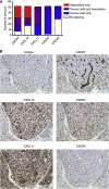
CXCR7 expression increases with glioma tumour grade. A multi-grade glioma array was stained by IHC with the CXCR7 mAb 11G8. (A) The incidence of 11G8 staining, on the tumour cells and/or tumour-associated vasculature, increased with tumour grade. (B) IHC staining of representative tumours with 11G8 (right column) or an isotype control mAb (left column). Note the staining of tumour cells (middle row) and tumour-associated vasculature (bottom row) in the Grade IV tumours by 11G8. Magnification × 200.
Figure 5
Figure 5
CXCR7 is expressed in the vicinity of its ligands in human GBM. GBM arrays were stained by IHC with antibodies specific for CXCR7, CXCR4, CXCR3, CXCL12 and CXCL11. (A) CXCR7, CXCL12 and CXCL11 were expressed on both tumour cells and tumour-associated vasculature, whereas CXCR4 and CXCR3 were expressed primarily only on tumour cells. (B) IHC staining of a representative GBM tumour with each antibody, as well as an isotype control antibody. In this tumour, CXCR7 is expressed primarily on the vasculature, CXCL12 and CXCL11 are expressed on the vasculature and tumour cells, CXCR4 is expressed primarily on tumour cells and CXCR3 is not expressed. Magnification × 200.
Figure 6
Figure 6
CCX662 increases post-IR survival time in rats containing C6 GBM tumours. C6 GBM cells were implanted intracranially into rats and, 7 days later, given 18 Gy whole-brain IR. Immediately following IR, rats were infused with CCX662 or its vehicle for 1 week. Surviving rats received another 1-week infusion of CCX662 or vehicle. CCX662 significantly increased post-IR median survival time (**P=0.0025 compared with vehicle, log-rank test; n=15–18 rats, compiled from two separate studies).
Figure 7
Figure 7
CXCR7 is expressed on the vasculature in C6 GBM tumours. (A) H&E staining of a representative tumour at × 20 magnification. (B) IHC staining of a representative tumour with the CXCR7 mAb 11G8 or an isotype control mAb at × 200 magnification.
Figure 8
Figure 8
CXCR7 is required for CSC activity in vitro. (A) The neurosphere formation assay using CXCR7bright and CXCR7dim sorted cells from GBM xenografts T3832, T387 and T4121. CXCR7bright cells exhibited a greater ability to form spheres than did CXCR7dim cells. (B) Neurosphere formation assay using sorted CD133+ CSCs from GBM xenograft T4121. CXCR7 inhibitor CCX771, but not its inactive chemical analogue CCX704, inhibited sphere formation. Inhibition of sphere formation was greater with 1 μM CCX771 than with 100 nM CX771 or with 1 μg ml−1 (1.3 μM) AMD3100. Similar results were observed with GBM xenograft T3832 (not shown).

Similar articles

Cited by

References

    1. Ahn GO, Brown JM. Matrix metalloproteinase-9 is required for tumor vasculogenesis but not for angiogenesis: role of bone marrow-derived myelomonocytic cells. Cancer Cell. 2008;13:193–205. - PMC - PubMed
    1. Ahn GO, Tseng D, Liao CH, Dorie MJ, Czechowicz A, Brown JM. Inhibition of Mac-1 (CD11b/CD18) enhances tumor response to radiation by reducing myeloid cell recruitment. Proc Natl Acad Sci USA. 2010;107:8363–8368. - PMC - PubMed
    1. Asahara T, Murohara T, Sullivan A, Silver M, van der Zee R, Li T, Witzenbichler B, Schatteman G, Isner JM. Isolation of putative progenitor endothelial cells for angiogenesis. Science. 1997;275:964–967. - PubMed
    1. Bao S, Wu Q, McLendon RE, Hao Y, Shi Q, Hjelmeland AB, Dewhirst MW, Bigner DD, Rich JN. Glioma stem cells promote radioresistance by preferential activation of the DNA damage response. Nature. 2006;444:756–760. - PubMed
    1. Berahovich RD, Wang Y, Jaen JC, Schall TJ. CXCR7 protein is not expressed on human or mouse leukocytes. J Immunol. 2010;185:5130–5139. - PubMed

Publication types

MeSH terms