Review
. 2014 Apr;35(2):159-94.
doi: 10.1210/er.2013-1087. Epub 2013 Dec 13.

Central regulation of hypothalamic-pituitary-thyroid axis under physiological and pathophysiological conditions

Affiliations
Review

Central regulation of hypothalamic-pituitary-thyroid axis under physiological and pathophysiological conditions

Csaba Fekete et al. Endocr Rev. 2014 Apr.

Abstract

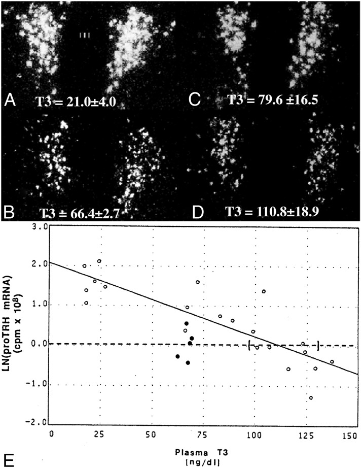
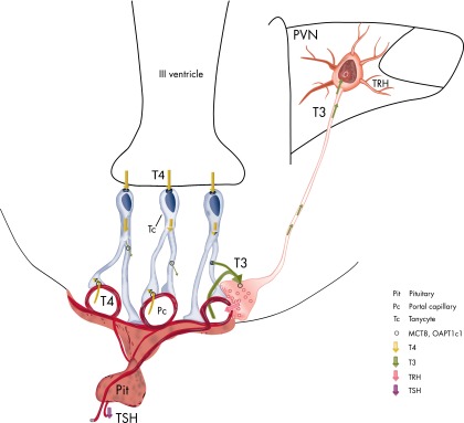
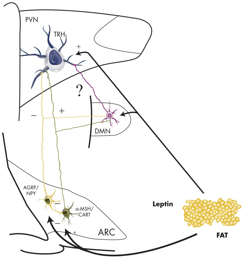
TRH is a tripeptide amide that functions as a neurotransmitter but also serves as a neurohormone that has a critical role in the central regulation of the hypothalamic-pituitary-thyroid axis. Hypophysiotropic TRH neurons involved in this neuroendocrine process are located in the hypothalamic paraventricular nucleus and secrete TRH into the pericapillary space of the external zone of the median eminence for conveyance to anterior pituitary thyrotrophs. Under basal conditions, the activity of hypophysiotropic TRH neurons is regulated by the negative feedback effects of thyroid hormone to ensure stable, circulating, thyroid hormone concentrations, a mechanism that involves complex interactions between hypophysiotropic TRH neurons and the vascular system, cerebrospinal fluid, and specialized glial cells called tanycytes. Hypophysiotropic TRH neurons also integrate other humoral and neuronal inputs that can alter the setpoint for negative feedback regulation by thyroid hormone. This mechanism facilitates adaptation of the organism to changing environmental conditions, including the shortage of food and a cold environment. The thyroid axis is also affected by other adverse conditions such as infection, but the central mechanisms mediating suppression of hypophysiotropic TRH may be pathophysiological. In this review, we discuss current knowledge about the mechanisms that contribute to the regulation of hypophysiotropic TRH neurons under physiological and pathophysiological conditions.


Figures

Figure 1.
Figure 1.
Schematic illustration of the organization of the rat preproTRH gene.
Figure 2.
Figure 2.
Distribution of TRH-synthesizing neurons in the rat PVN. A–C, Low-power micrographs illustrate the TRH neurons at three rostrocaudal levels of the PVN. D–F, Schematic drawings illustrate the subdivisions of the PVN where hypophysiotropic TRH neurons are localized (gray). AP, Anterior parvocellular subdivision; DP, dorsal parvocellular subdivision; LP, lateral parvocellular subdivision; MN, magnocellular part of PVN; MP, medial parvocellular subdivision, PV, periventricular parvocellular subdivision; VP, ventral parvocellular subdivision; III, third ventricle. [Reproduced from C. Fekete and R. M. Lechan: Negative feedback regulation of hypophysiotropic thyrotropin-releasing hormone (TRH) synthesizing neurons: role of neuronal afferents and type 2 deiodinase. Front Neuroendocrinol. 2007;28:97–114 (58), with permission. © Elsevier.]
Figure 3.
Figure 3.
Darkfield photomicrographs showing proTRH mRNA expression in the anterior, mid, and posterior levels of the PVN in control (A–C) and hypothyroid (D–F) rats and in control (G–I) and hypothyroid (J–L) mice. Note the dramatic increase in silver grains denoting proTRH mRNA in the mid and caudal level of the hypothyroid rat PVN (E and F), whereas hypothyroidism increases proTRH mRNA only in midlevel neurons in mice (K). III, Third ventricle. [Panels G–L were reproduced from A. Kádár et al: Distribution of hypophysiotropic thyrotropin-releasing hormone (TRH)-synthesizing neurons in the hypothalamic paraventricular nucleus of the mouse. J Comp Neurol. 2010;518:3948–3961 (44), with permission. © Wiley-Liss Inc.]
Figure 4.
Figure 4.
Distribution of TRH-IR terminals in the mouse median eminence. TRH-IR axons densely innervate the external zone of the median eminence. III, Third ventricle; ME, median eminence.
Figure 5.
Figure 5.
Innervation of the TRH neurons in the rat PVN by axons originating from the arcuate nucleus (A), DMN (B), and catecholaminergic neurons (C) in the brainstem. A, TRH neurons (blue) are contacted by axon terminals containing α-MSH (red; arrowhead) and AGRP (green; arrows). B, Axon varicosities containing the anterogradely transported marker protein, PHA-L (black) are juxtaposed to TRH-synthesizing neurons (brown) after iontophoretic administration of the tracer into the DMN. C, Both noradrenergic (red; open arrows) and adrenergic (yellow, white arrows) axons establish contacts with the TRH neurons. [Modified from C. Fekete et al: α-Melanocyte-stimulating hormone is contained in nerve terminals innervating thyrotropin-releasing hormone-synthesizing neurons in the hypothalamic paraventricular nucleus and prevents fasting-induced suppression of prothyrotropin-releasing hormone gene expression. J Neurosci. 2000;20:1550–1558 (52), with permission. © Society for Neuroscience. From E. Mihály et al: Hypothalamic dorsomedial nucleus neurons innervate thyrotropin-releasing hormone-synthesizing neurons in the paraventricular nucleus. Brain Res. 2001;891:20–31 (74), with permission. © Elsevier. And from T. Füzesi et al: Noradrenergic innervation of hypophysiotropic thyrotropin-releasing hormone-synthesizing neurons in rats. Brain Res. 2009;1294:38–44 (61), with permission. © Elsevier.]
Figure 6.
Figure 6.
Schematic drawing summarizing known inputs to TRH neurons in the PVN. The inputs with identified origins are depicted by a neuron sending its axon to the TRH neuron. Inputs with currently unknown origins are labeled with axon terminals on the surface of the TRH neuron. PACAP, Pituitary adenylate cyclase-activating polypeptide.
Figure 7.
Figure 7.
A and B, Organization of tanycyte subtypes in the MBH. A, Vimentin-immunolabeled (red) coronal section with DAPI counterstaining (blue) shows the distribution of tanycytes and their processes. B, The schematic diagram illustrates the location of tanycyte subtypes in the wall and floor of the third ventricle. C and D, All tanycyte subtypes synthesize MCT8 (C) and OATP1C1 (D) thyroid hormone transporters. E, Silver grains denoting D2 mRNA are accumulated over the cells lining the wall of the third ventricle, the tuberoinfundibular sulci (arrowheads), and around blood vessels in the arcuate nucleus (arrows). F and G, Higher power micrographs show the association of D2 mRNA with the tuberoinfundibular sulcus (arrowheads) and a blood vessel (arrows) in the arcuate nucleus (F), and in the external zone of the median eminence (G). H, Tanycyte expression of PPII mRNA. III, Third ventricle; Arc, arcuate nucleus; ME, median eminence; VMN, ventromedial nucleus. [Modified from E. Sánchez et al: Tanycyte pyroglutamyl peptidase II contributes to regulation of the hypothalamic-pituitary-thyroid axis through glial-axonal associations in the median eminence. Endocrinology. 2009;150:2283–2291 (35), with permission. © The Endocrine Society. From Kalló et al: A novel pathway regulates thyroid hormone availability in rat and human hypothalamic neurosecretory neurons. PLoS One. 2012;7:e37860 (51), with permission. © Public Library of Science. And from C. Fekete et al: DARPP-32 and CREB are present in type 2 iodothyronine deiodinase-producing tanycytes: implications for the regulation of type 2 deiodinase activity. Brain Res. 2000;862:154–161 (124), with permission. © Elsevier. Courtesy of Dr Gábor Wittmann.]
Figure 8.
Figure 8.
In situ hybridization autoradiograms showing the effect of hypo- and hyperthyroidism on proTRH mRNA level in the medial parvocellular subdivision of the PVN. A substantial increase in silver grain accumulation is observed in the hypothyroid animal (A) compared to the fed control (B). In contrast, hyperthyroidism results in a marked reduction of proTRH mRNA level in the PVN (C). [Modified from E. M. Dyess et al: Triiodothyronine exerts direct cell-specific regulation of thyrotropin-releasing hormone gene expression in the hypothalamic paraventricular nucleus. Endocrinology. 1988;123:2291–2297 (115), with permission. © The Endocrine Society. And from C. Fekete and R. M. Lechan: Negative feedback regulation of hypophysiotropic thyrotropin-releasing hormone (TRH) synthesizing neurons: role of neuronal afferents and type 2 deiodinase. Front Neuroendocrinol. 2007;28:97–114 (58), with permission. © Elsevier.]
Figure 9.
Figure 9.
In situ hybridization autoradiographs of proTRH mRNA in the PVN of hypothyroid (A), euthyroid (B), and hypothyroid (C and D) animals receiving a constant infusion of 0.5 μg (C) or 0.75 μg (D) of T3 per 100 g body weight per day. Mean plasma T3 levels (±SEM) are shown for each group at the bottom of the photomicrographs. Note that only the higher dose of T3 that raised plasma T3 levels into the supranormal range was capable of suppressing proTRH mRNA to euthyroid levels. E, Regression analysis of the above experiment. Interrupted line represents the mean ln(proTRH mRNA) for euthyroid animals, and its intercept with the regression line estimates the plasma T3 concentration required to suppress proTRH mRNA to euthyroid levels. Ninety-five percent confidence intervals for each intercept are bracketed. Open dots denote values for hypothyroid animals and hypothyroid animals infused with graded doses of T3. Closed dots denote values for euthyroid controls. [Modified from I Kakucska et al: Thyrotropin-releasing hormone gene expression in the hypothalamic paraventricular nucleus is dependent upon feedback regulation by both triiodothyronine and thyroxine. Endocrinology. 1992;130:2845–2850 (123), with permission. © The Endocrine Society. And from C. Fekete and R. M. Lechan: Negative feedback regulation of hypophysiotropic thyrotropin-releasing hormone (TRH) synthesizing neurons: role of neuronal afferents and type 2 deiodinase. Front Neuroendocrinol. 2007;28:97–114 (58), with permission. © Elsevier.]
Figure 10.
Figure 10.
Schematic illustration of the machinery involved in negative feedback regulation of the HPT axis by thyroid hormone.
Figure 11.
Figure 11.
Darkfield illumination photomicrographs of proTRH mRNA in the hypothalamic PVN in (A) fed, (B) fasted, and (C) fasted animals receiving leptin. Note the marked reduction in silver grains over neurons in the PVN in the fasted animals but restoration to normal in the fasted animals receiving leptin. III, Third ventricle. [Reproduced from C. Fekete and R. M. Lechan: Negative feedback regulation of hypophysiotropic thyrotropin-releasing hormone (TRH) synthesizing neurons: role of neuronal afferents and type 2 deiodinase. Front Neuroendocrinol. 2007;28:97–114 (58), with permission. © Elsevier.]
Figure 12.
Figure 12.
Schematic drawing summarizing the regulation of TRH neurons by fasting.
Figure 13.
Figure 13.
Darkfield illumination micrographs of proTRH mRNA in the medial and periventricular parvocellular subdivisions of the hypothalamic PVN in fed animals (A and D), fasted animals (B and E), and fasted animals receiving an intracerebroventricular infusion of either α-MSH (C) or CART (F) every 6 hours for 64 hours. Note the reduction in the accumulation of silver grains over the PVN in fasted animals compared with the fed controls. Both α-MSH and CART administration prevent the fasting-induced fall of proTRH mRNA. III, Third ventricle. [Modified from C. Fekete et al: Association of cocaine- and amphetamine-regulated transcript-immunoreactive elements with thyrotropin-releasing hormone-synthesizing neurons in the hypothalamic paraventricular nucleus and its role in the regulation of the hypothalamic-pituitary-thyroid axis during fasting. J Neurosci. 2000;20:9224–9234 (42), with permission. © Society for Neuroscience. From C. Fekete et al: α-Melanocyte-stimulating hormone is contained in nerve terminals innervating thyrotropin-releasing hormone-synthesizing neurons in the hypothalamic paraventricular nucleus and prevents fasting-induced suppression of prothyrotropin-releasing hormone gene expression. J Neurosci. 2000;20:1550–1558 (52), with permission. © Society for Neuroscience. And from C. Fekete and R. M. Lechan: Negative feedback regulation of hypophysiotropic thyrotropin-releasing hormone (TRH) synthesizing neurons: role of neuronal afferents and type 2 deiodinase. Front Neuroendocrinol. 2007;28:97–114 (58), with permission. © Elsevier.]
Figure 14.
Figure 14.
Darkfield illumination photomicrographs of proTRH mRNA in the medial parvocellular subdivision of the PVN in control (A), AGRP-treated (B), and NPY-treated (C) animals. Note the marked reduction in silver grains over neurons in the PVN in both the AGRP- and NPY-infused groups. III, Third ventricle. [Modified from C. Fekete et al: Agouti-related protein (AGRP) has a central inhibitory action on the hypothalamic-pituitary-thyroid (HPT) axis; comparisons between the effect of AGRP and neuropeptide Y on energy homeostasis and the HPT axis. Endocrinology. 2002;143:3846–3853 (155), with permission. © The Endocrine Society. And from C. Fekete and R. M. Lechan: Negative feedback regulation of hypophysiotropic thyrotropin-releasing hormone (TRH) synthesizing neurons: role of neuronal afferents and type 2 deiodinase. Front Neuroendocrinol. 2007;28:97–114 (58), with permission. © Elsevier.]
Figure 15.
Figure 15.
A and B, Regulation of D2 gene expression in tanycytes by LPS. Low-power darkfield micrographs of the caudal part of the hypothalamus show the D2 expression in control animals (A) and 12 hours after LPS administration (B). Note marked increase in the density of silver grains, particularly in the external zone of the median eminence. C–E, Darkfield illumination photomicrographs of Iκ-Bα mRNA expression in the MBH in animals receiving saline (C) or LPS 3 hours (D) or 12 hours (E) before sacrifice. Note the marked accumulation of silver grains over the pars tuberalis in panel D 3 hours after the administration of LPS. Iκ-Bα mRNA is only seen in a subset of α tanycytes 12 hours after LPS administration (arrows in panel E). F and G (arrows), Expression of TSHβ mRNA in the pars tuberalis of animals receiving saline (F) or LPS (G) 9 hours before sacrifice. Note increased expression of TSHβ mRNA in the PT after LPS administration. Ventricular borders are demarcated with dotted lines. H, Double-labeled fluorescent image illustrates the presence of phospho-CREB (green and yellow, arrows) in the nucleus of tanycytes (red) 9 hours after administration of LPS. III, Third ventricle; ME, median eminence; PT, pars tuberalis. [Modified from C. Fekete et al: Lipopolysaccharide induces type 2 iodothyronine deiodinase in the mediobasal hypothalamus: implications for the nonthyroidal illness syndrome. Endocrinology. 2004;145:1649–1655 (99), with permission. © The Endocrine Society. And from E. Sánchez et al: Contribution of TNF-α and nuclear factor-κB signaling to type 2 iodothyronine deiodinase activation in the mediobasal hypothalamus after lipopolysaccharide administration. Endocrinology. 2010;151:3827–3835 (142), with permission. © The Endocrine Society.]

References

    1. Lechan RM, Hollenberg A, Fekete C. Hypothalamic-pituitary-thyroid axis: organization, neural/endocrine control of TRH. In: Squire LR, ed. Encyclopedia of Neuroscience. Oxford, UK: Academic Press; 2009:75–87
    1. Reichlin S. TRH: historical aspects. Ann NY Acad Sci. 1989;553:1–6 - PubMed
    1. Boler J, Enzmann F, Folkers K, Bowers CY, Schally AV. The identity of chemical and hormonal properties of the thyrotropin releasing hormone and pyroglutamyl-histidyl-proline amide. Biochem Biophys Res Commun. 1969;37:705–710 - PubMed
    1. Burgus R, Dunn TF, Desiderio D, Guillemin R. [Molecular structure of the hypothalamic hypophysiotropic TRF factor of ovine origin: mass spectrometry demonstration of the PCA-His-Pro-NH2 sequence]. C R Acad Sci Hebd Seances Acad Sci D. 1969;269:1870–1873 - PubMed
    1. Hashimoto K, Zanger K, Hollenberg AN, Cohen LE, Radovick S, Wondisford FE. cAMP response element-binding protein-binding protein mediates thyrotropin-releasing hormone signaling on thyrotropin subunit genes. J Biol Chem. 2000;275:33365–33372 - PubMed

Publication types

MeSH terms

Substances