Review
. 2014 Mar:253:197-207.
doi: 10.1016/j.expneurol.2013.12.024. Epub 2014 Jan 11.

Functional regeneration beyond the glial scar

Affiliations
Review

Functional regeneration beyond the glial scar

Jared M Cregg et al. Exp Neurol. 2014 Mar.

Abstract

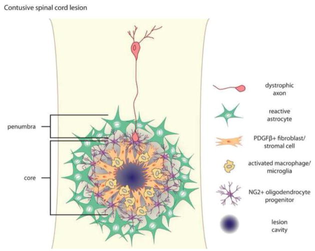
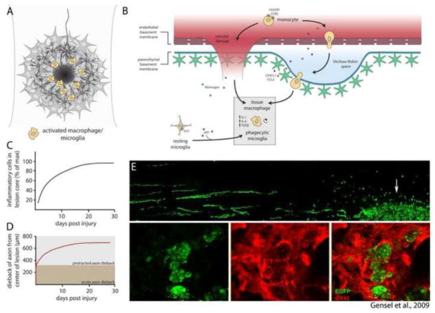
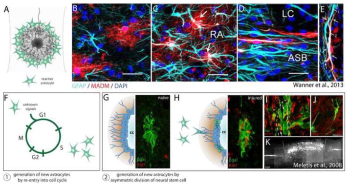
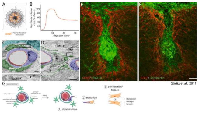
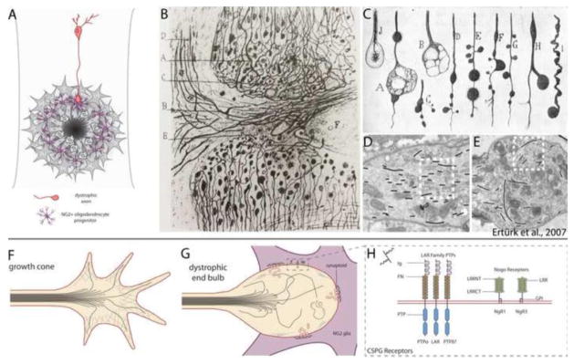
Astrocytes react to CNS injury by building a dense wall of filamentous processes around the lesion. Stromal cells quickly take up residence in the lesion core and synthesize connective tissue elements that contribute to fibrosis. Oligodendrocyte precursor cells proliferate within the lesion and entrap dystrophic axon tips. Here we review evidence that this aggregate scar acts as the major barrier to regeneration of axons after injury. We also consider several exciting new interventions that allow axons to regenerate beyond the glial scar, and discuss the implications of this work for the future of regeneration biology.

Keywords: Axon growth cone; Chondroitin sulfate proteoglycans; Glial scar; Hypertrophy; Regeneration; Spinal cord injury.


Figures

Fig. 1
Fig. 1
Anatomy of a contusive spinal cord lesion. Spinal cord lesions have two distinct components—the lesion penumbra is composed of hypertrophic astrocytes, and the lesion core is composed of NG2+ oligodendrocyte precursor cells, PDGFRβ+ fibroblasts, and macrophages/microglia. Dystrophic axons become entrapped within the lesion in close association with NG2 glia. The layered architecture of the glial scar is thought to reflect both the dynamic polarization of different populations of cells at distinct times after injury and segregation of distinct populations via chemorepulsion.
Fig. 2
Fig. 2
Inflammatory processes within the lesion core. (A) After injury, macrophages and microglia accumulate within the lesion core. (B) Recruitment of inflammatory cells occurs by extravasation of leukocytes from damaged blood vessels and migration of resident microglia to sites of CNS injury. Tissue macrophages and phagocytic microglia synthesize a contingent of cytokines that promote inflammation. (C) Accumulation of inflammatory cells within the lesion core reaches peak density by 30 days after injury (data adapted from Horn et al., 2008; Kigerl et al., 2009). (D) Dieback of injured axons occurs in two distinct phases: Acute axonal degeneration occurs via intracellular Ca2+-dependent cysteine proteases, whereas protracted axonal dieback occurs via direct interaction with inflammatory cells. Protracted axonal dieback correlates well with the accumulation of inflammatory cells within the lesion core (C). Data in (D) adapted from Kerschensteiner et al. (2005) and Horn et al. (2008). (E) Data republished from Gensel et al. (2009) with permission from the Society for Neuroscience; permission conveyed through the Copyright Clearance Center, Inc. EGFP labeled dorsal root ganglion neurons were microtransplanted at a site distant to zymosan injection. EGFP+ axons are observed growing toward the site of zymosan injection, where activated OX42+ macrophages (red) are observed engulfing DRG axon fragments.
Fig. 3
Fig. 3
Astrocyte heterogeneity. (A) Astrocytes become hypertrophic in response to CNS insult, forming a dense wall of filamentous processes at the lesion penumbra. (B–E) Data republished from Wanner et al. (2013) with permission from the Society for Neuroscience; permission conveyed through the Copyright Clearance Center, Inc. A GFAP-Cre/MADM-reporter genetic mosaic mouse was used to sparsely label astrocytes with RFP, enabling visualization of fine astrocytic processes, independent astrocytic domains, and astrocytes only weakly immunoreactive for GFAP. (B) In the uninjured cord astrocytes occupy mutually exclusive domains, exhibit a bushy morphology with many fine processes, and express varying amounts of the intermediate filament protein GFAP. (C) 14 days after injury, reactive astrocytes (RA) more distal to the lesion core exhibit increased expression of GFAP, however independent domains and stellate morphology are largely maintained. (D) At the lesion penumbra (ASB), astrocytes no longer maintain a bushy appearance, but take on an elongated morphology with extensive overlap of individual territory. (E) Two different astrocytes (1, 2) form a mesh-like layer of entangled filamentous processes at the lesion penumbra. (F–K) Generation of new astrocytes after spinal cord injury results from at least two distinct mechanisms: 1) Generation of new astrocytes by re-entry of adult astrocytes into the cell cycle. 2) Generation of new astrocytes by asymmetric division of neural stem cells lining the central canal. (G–K) Fluorescent images reprinted from Meletis et al. (2008). (G) Upon tamoxifen administration, FoxJ1-CreER drives βgal reporter expression in a population of ependymal cells lining the central canal. These cells undergo only basal levels of division in the uninjured cord (note absence of Ki67+ cells). (H) After injury, several βgal+ ependymal cells undergo division, demonstrated by co-expression with the proliferation marker Ki67. (I) βgal+ neural stem cell progeny migrate away from the ependymal cell layer lining the central canal (marked by a dashed line), a majority of which become astrocytes (J). (K) Sagittal section demonstrating that βgal+ neural stem cell progeny form a major component of the glial scar one month after injury. Note the specificity of the FoxJ1-CreER line, where βgal expression is confined to the central canal of the spinal cord in regions rostral and caudal to the lesion.
Fig. 4
Fig. 4
Fibrosis within a contusive lesion. (A) Fibroblasts take up residence within the lesion core, reaching their maximum density around 10 days post injury (B, data adapted from Göritz et al., 2011; Soderblom et al., 2013). (C–F) Data reprinted from Göritz et al. (2011) with permission from AAAS. (C) Electron micrograph of a perivascular niche 5 days post injury. Whereas macrostructure is largely maintained, Type-A pericytes (pseudocolored green) have detached from the surrounding basal lamina (bl) and deposited ECM within the basal lamina sheath. (D) 14 days after injury several type-A pericytes (green) have left their perivascular niche and exhibit extensive fibrosis. (E) 18 weeks post injury the lesion core is occupied by a large contingent of PDGFβ+ fibroblasts, which are associated with deposition of fibronectin in the lesion core (F). (G) Fibrosis within a contusive lesion is caused by perivascular progenitors, and occurs in three distinct phases: 1) Injury causes delamination of pericytes from the endothelial basement membrane. 2) Pericytes that have detached from the basement membrane undergo transition into a mesenchymal state. 3) Progenitors give rise to fibroblasts, which synthesize extracellular matrix that contributes to fibrosis.
Fig. 5
Fig. 5
The glial scar acts as the primary barrier to regenerating axons. (A) Regenerating axons halt abruptly at the glial scar in close association with NG2 glia, and axon endings take on a state of dystrophy. (B, C) Reprinted from Cajal’s Degeneration and Regeneration of the Nervous System (1928) by permission of Oxford University Press, USA. (B) Drawing by Ramón y Cajal of the edges of a complete transection lesion to the spinal cord. Cajal noted that adjacent to the lesion, fine axons terminate in rings or little clubs while larger axons end in voluminous clubs. (C) Several drawings by Cajal of the various appearances of retraction clubs at the lesion. (D, E) Data republished from Ertürk et al. (2007) with permission from the Society for Neuroscience; permission conveyed through the Copyright Clearance Center, Inc. (D) Electron micrograph of a growth cone in the lesioned sciatic nerve. Growth cones exhibit highly parallel arrays of microtubules (traced with black lines). (E) Electron micrograph of a retraction bulb from a lesioned central axon of a dorsal root ganglion neuron. Retraction bulbs exhibit disorganized or splayed microtubules. (F) Illustration of a growth cone. Microtubules are bundled and oriented toward the direction of the growing axon. Growth cones also have a highly organized F-actin network and several filopodia. (G) Dystrophic end bulbs have disorganized microtubules and a disrupted F-actin network. Large membrane blebs or inclusions can be observed within retraction clubs. Dystrophic axons associate with NG2 glia in the outer margin of the lesion core, forming “synaptoids” that exhibit similar properties to a mature synapse, with numerous presynaptic vesicles and omega structures, and a post-synaptic density at the NG2 glial membrane. (H) Several recently identified CSPG receptors act to signal inhibition, and may entrap axons on the surface of NG2 glia. These receptors fall into two classes: the LAR family of transmembrane protein tyrosine phosphatases (which includes PTPσ, LAR, and PTPδ), and the Nogo receptors NgR1 and NgR3. LAR family receptor protein tyrosine phosphatases have high sequence similarity and contain three N-terminal Ig domains, 8 fibronectin repeats, and two intracellular tandem phosphatase domains. The first Ig domain of PTPσ, LAR, and PTPδ contains a canonical GAG binding motif (BB-X-BB, where B is lysine or arginine). NgR1 and NgR3 have several leucine-rich repeats, are GPI-anchored, and contain a cluster of basic residues in the C-terminal stalk required for binding CSPGs.

References

    1. Ajami B, Bennett JL, Krieger C, McNagny KM, Rossi FM. Infiltrating monocytes trigger EAE progression, but do not contribute to the resident microglia pool. Nat Neurosci. 2011;14:1142–1149. - PubMed
    1. Alilain WJ, Horn KP, Hu H, Dick TE, Silver J. Functional regeneration of respiratory pathways after spinal cord injury. Nature. 2011;475:196–200. - PMC - PubMed
    1. Alto LT, Havton LA, Conner JM, Hollis ER, II, Blesch A, Tuszynski M. Chemotropic guidance facilitates axonal regeneration and synapse formation after spinal cord injury. Nat Neurosci. 2009;12:1106–1113. - PMC - PubMed
    1. Aricescu AR, McKinnell IW, Halfter W, Stocker AW. Heparan sulfate proteoglycans are ligands for receptor protein tyrosine phosphatase sigma. Mol Cell Biol. 2002;22:1881–1892. - PMC - PubMed
    1. Asher RA, Morgenstern DA, Fidler PS, Adcock KH, Oohira A, Braistead JE, Levine JM, Margolis RU, Rogers JH, Fawcett JW. Neurocan is upregulated in the injured brain and in cytokine-treated astrocytes. J Neurosci. 2000;20:2427–2438. - PMC - PubMed

Publication types