. 2014 Jan 14;15(1):1003-13.
doi: 10.3390/ijms15011003.

Lactoferrin directly scavenges hydroxyl radicals and undergoes oxidative self-degradation: a possible role in protection against oxidative DNA damage

Affiliations

Lactoferrin directly scavenges hydroxyl radicals and undergoes oxidative self-degradation: a possible role in protection against oxidative DNA damage

Yuki Ogasawara et al. Int J Mol Sci. .

Abstract

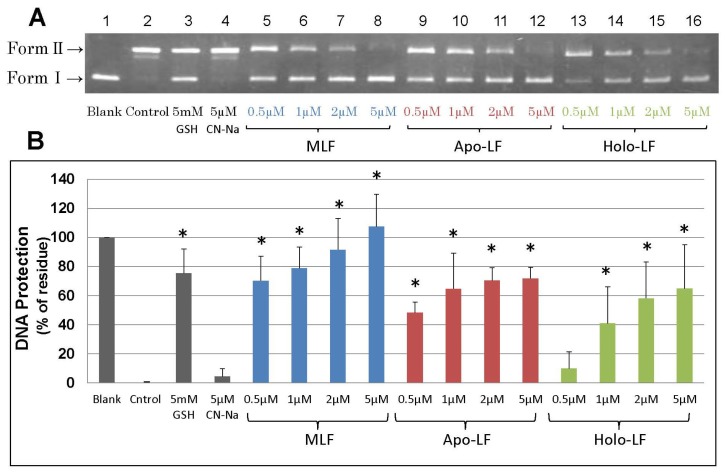
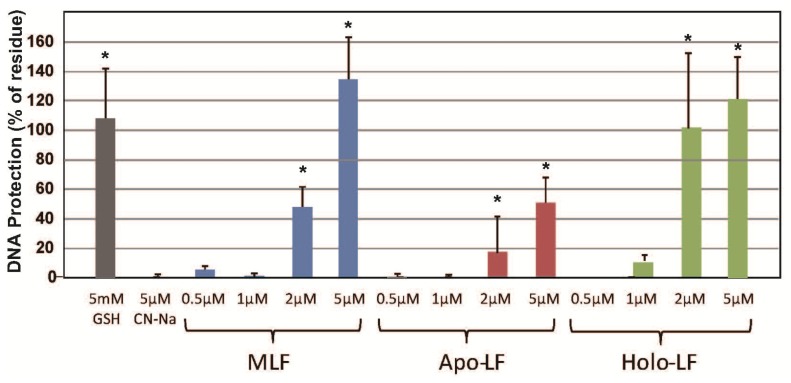
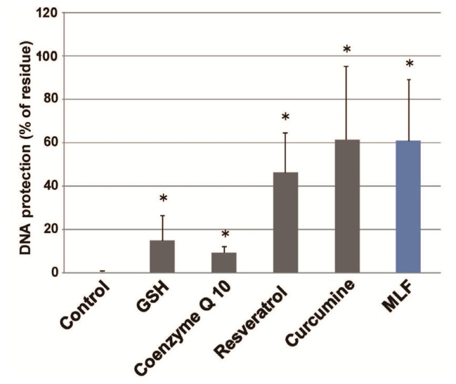
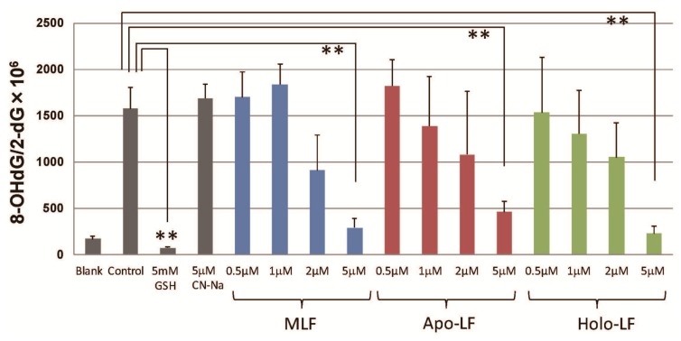
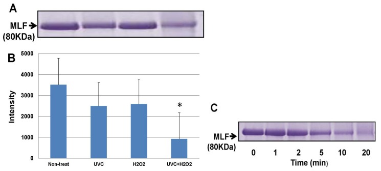
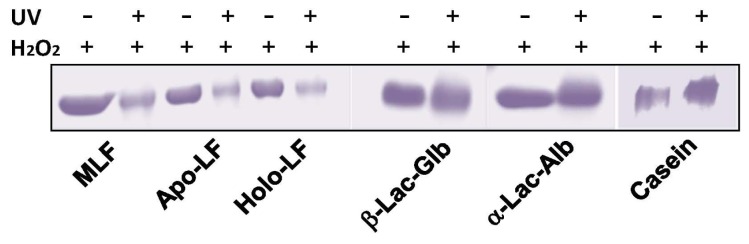
In this study, we examined the protective effect of lactoferrin against DNA damage induced by various hydroxyl radical generation systems. Lactoferrin (LF) was examined with regard to its potential role as a scavenger against radical oxygen species using bovine milk LF. Native LF, iron-saturated LF (holo-LF), and apolactoferrin (apo-LF) effectively suppressed strand breaks in plasmid DNA due to hydroxyl radicals produced by the Fenton reaction. In addition, both native LF and holo-LF clearly protected calf thymus DNA from fragmentation due to ultraviolet irradiation in the presence of H2O2. We also demonstrated a protective effect of all three LF molecules against 8-hydroxydeoxyguanosine (8-OHdG) formation in calf thymus DNA following ultraviolet (UV) irradiation with H2O2. Our results clearly indicate that native LF has reactive oxygen species-scavenging ability, independent of its nature as a masking component for transient metals. We also demonstrated that the protective effect of LF against oxidative DNA damage is due to degradation of LF itself, which is more susceptible to degradation than other bovine milk proteins.


Figures

Figure 1.
Figure 1.
Dose response and efficacy of LFs on DNA damage by •OH generated by the Fenton reaction. Electrophoresis of plasmid DNA using an agarose gel (1.0%) was performed after exposure to •OH generated by the Fenton reaction. Experiments were conducted for 20 min at 37 °C, using iron and H2O2 (using final concentrations of 50 μL PBS, 50 μM H2O2, 5 μM FeCl3, 25 μM EDTA, and 10 μM ascorbic acid). (A) Lane 1, plasmid (Blank); lane 2, Fenton reaction mixture plus plasmid (Control); lane 3, Fenton reaction mixture plus plasmid and 5 mM GSH; lane 4, Fenton reaction mixture plus plasmid and 5 μM Casein sodium (CN-Na); lane 5, Fenton reaction mixture plus plasmid and 0.5 μM MLF; lane 6, Fenton reaction mixture plus plasmid and 1 μM MLF; lane 7, Fenton reaction mixture plus plasmid and 2 μM MLF; lane 8, Fenton reaction mixture plus plasmid and 5 μM MLF; lane 9, Fenton reaction mixture plus plasmid and 0.5 μM apo-LF; lane 10, Fenton reaction mixture plus plasmid and 1 μM apo-LF; lane 11, Fenton reaction mixture plus plasmid and 2 μM apo-LF; lane 12, Fenton reaction mixture plus plasmid and 5 μM apo-LF; lane 13, Fenton reaction mixture plus plasmid and 0.5 μM holo-LF; lane 14, Fenton reaction mixture plus plasmid and 1 μM holo-LF; lane 15, Fenton reaction mixture plus plasmid and 2 μM holo-LF; and lane 16, Fenton reaction mixture plus plasmid and 5 μM holo-LF; (B) DNA protection (%) was calculated based on the densitometry of EtBr-stained bands (Form I) against blank (non-treated plasmid DNA, lane 1) band intensities under the reaction conditions described in A (lanes 2–16). Data are presented as the mean ± S.D. of triplicate determinations. * p < 0.05 compared to the control value was considered as a statistically significant difference.
Figure 2.
Figure 2.
Dose responses and efficacy of LFs on calf thymus DNA strand breaks by UV irradiation in the presence of H2O2. Electrophoresis of calf thymus DNA using an agarose gel (1.0%) was performed following exposure to UV (254 nm) irradiation with 5 mM H2O2. Reactions were conducted for 10 min at room temperature. DNA protection (%) was calculated based on the densitometry of EtBr-stained bands vs. a non-treated sample (Control). Data are presented as the mean ± S.D. of triplicate determinations. * p < 0.05 compared to the CN-Na (negative control) value was considered as a statistically significant difference.
Figure 3.
Figure 3.
Protective effects of LFs and various antioxidants on calf thymus DNA strand breaks of p following exposure to •OH generated by the UV-H2O2 system. The effects of 5 μM MLF and various other compounds (5 mM GSH, 50 μM resveratorol, 50 μM curcumine, and 50 μM Coenzyme Q10) were determined by electrophoresis of DNA. Electrophoresis of calf thymus DNA using agarose gel (1.0%) was performed following exposure to UV irradiation (254 nm) with 5 mM H2O2 in the presence of various test compounds. Reactions were conducted for 10 min at room temperature. DNA protection (%) was calculated based on the densitometry of EtBr-stained bands vs. control band intensities. Data are presented as the mean ± S.D. of triplicate determinations. * p < 0.05 compared to the control value was considered as a statistically significant difference.
Figure 4.
Figure 4.
Effects of LFs on 8-OHdG formation following exposure to •OH generated by the UV-H2O2 system. 8-OHdG formation in calf thymus DNA following UV irradiation (254 nm) in the presence of H2O2 was determined as described in the Materials and Methods Section. Reactions with or without LFs were conducted for 5 min at room temperature. Data are presented as the mean ± S.D. of triplicate determinations. ** p < 0.01 compared to the control value obtained was considered as a statistically significant difference.
Figure 5.
Figure 5.
SDS gel electrophoresis of LF and apo-LF solutions exposed to UV irradiation with H2O2. (A) CBB stained for native LF (MLF) in SDS-polyacrylamide gel. Lane 1, non-treated; lane 2, UV (254 nm) irradiated for 10 min without H2O2; lane 3, H2O2-treated without UV irradiation; and lane 4, UV irradiated for 10 min with H2O2; (B) Densitometry of the stained bands demonstrated that 80-kDa native LF (MLF) remains intact under the conditions described in (A). Data are presented as the mean ± S.D. of triplicate determinations. * p < 0.05 compared to the non-treated control values obtained was considered as a statistically significant difference; (C) Coomassie brilliant blue (CBB) stained in SDS-polyacrylamide gel for native LF (MLF) exposed to UV (254 nm) irradiation with H2O2 for different lengths of time. Lanes from left to right: 0, 1, 2, 5, 10 and 20 min.
Figure 6.
Figure 6.
Degradation of LFs and other milk proteins exposed to UV irradiation-induced hydroxyl radicals. CBB stained for native LF (MLF), apo-LF, holo-LF, β-lactogloblin (Lac-Glb), and α-lactoalbumin (Lac-Alb), in SDS-polyacrylamide gel (5%–20%). Each protein was treated with or without UV-irradiation in the presence of 5 mM H2O2 for 10 min.

References

    1. Schanbacher F.L., Goodman R.E., Talhouk R.S. Bovine mammary lactoferrin: Implications from messenger ribonucleic acid (mRNA) sequence and regulation contrary to other milk proteins. J. Dairy. Sci. 1993;76:3812–3831. - PubMed
    1. van der Strate B.W., Beljaars L., Molema G., Harmsen M.C., Meijer D.K. Antiviral activities of lactoferrin. Antiviral Res. 2001;52:225–239. - PubMed
    1. Bennett R.M., Kokocinski T. Lactoferrin content of peripheral blood cells. Br. J. Haematol. 1978;39:509–521. - PubMed
    1. Caccavo D., Pellegrino N.M., Altamura M., Rigon A., Amati L., Amoroso A., Jirillo E. Antimicrobial and immunoregulatory functions of lactoferrin and its potential therapeutic application. J. Endotoxin Res. 2002;8:403–417. - PubMed
    1. Conneely O.M. Antiinflammatory activities of lactoferrin. J. Am. Coll. Nutr. 2001;20:389S–395S. - PubMed

MeSH terms

LinkOut - more resources