. 2014 Apr;34(4):660-7.
doi: 10.1038/jcbfm.2013.242. Epub 2014 Jan 15.

NLRP3 deficiency ameliorates neurovascular damage in experimental ischemic stroke

Affiliations

NLRP3 deficiency ameliorates neurovascular damage in experimental ischemic stroke

Fan Yang et al. J Cereb Blood Flow Metab. 2014 Apr.

Abstract

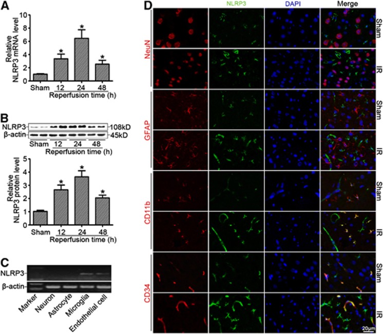
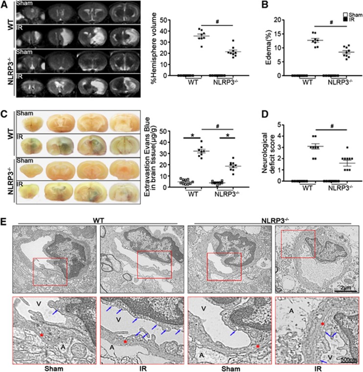
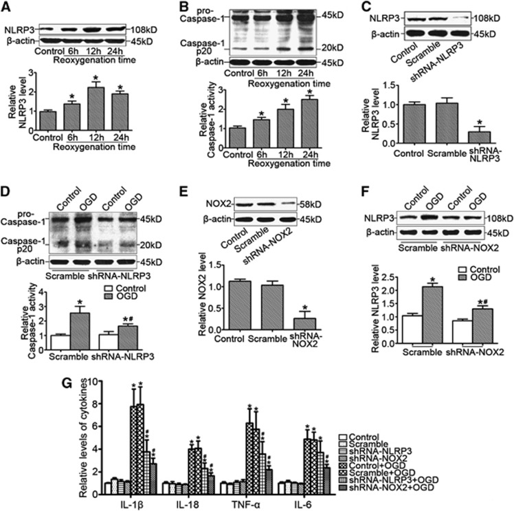
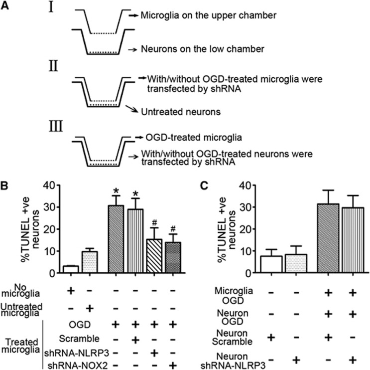
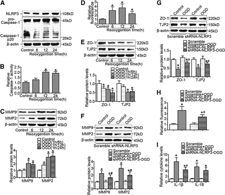
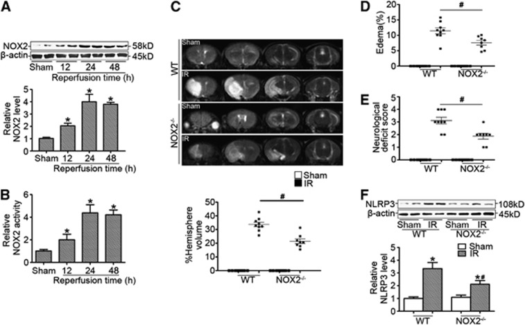
Although the innate immune response to induce postischemic inflammation is considered as an essential step in the progression of cerebral ischemia injury, the role of innate immunity mediator NLRP3 in the pathogenesis of ischemic stroke is unknown. In this study, focal ischemia was induced by middle cerebral artery occlusion in NLRP3(-/-), NOX2(-/-), or wild-type (WT) mice. By magnetic resonance imaging (MRI), Evans blue permeability, and electron microscopic analyses, we found that NLRP3 deficiency ameliorated cerebral injury in mice after ischemic stroke by reducing infarcts and blood-brain barrier (BBB) damage. We further showed that the contribution of NLRP3 to neurovascular damage was associated with an autocrine/paracrine pattern of NLRP3-mediated interleukin-1β (IL-1β) release as evidenced by increased brain microvessel endothelial cell permeability and microglia-mediated neurotoxicity. Finally, we found that NOX2 deficiency improved outcomes after ischemic stroke by mediating NLRP3 signaling. This study for the first time shows the contribution of NLRP3 to neurovascular damage and provides direct evidence that NLRP3 as an important target molecule links NOX2-mediated oxidative stress to neurovascular damage in ischemic stroke. Pharmacological targeting of NLRP3-mediated inflammatory response at multiple levels may help design a new approach to develop therapeutic strategies for prevention of deterioration of cerebral function and for the treatment of stroke.


Figures

Figure 1
Figure 1
NLRP3 expression was significantly increased after cerebral ischemia reperfusion (IR). (A) Relative mRNA levels of NLRP3 by real-time RT-PCR analysis in the ischemic cerebral hemisphere from wild-type (WT) mice at different time points after reperfusion. (B) Western blot analysis of NLRP3 protein levels in the ischemic cerebral hemisphere from WT mice at different time points after reperfusion. (C) NLRP3 mRNA detection by RT-PCR from primary cultured cortical neurons, astrocytes, microglia, and brain microvessel endothelial cells in vitro. (D) Cellular localization of NLRP3 at 24 hours of reperfusion after middle cerebral artery occlusion (MCAO) showing that NLRP3 was expressed in microglia and endothelial cells rather than in astrocytes and neurons of ischemic brains by confocal analysis. *P<0.05 vs. sham-operated mice (n=8).
Figure 2
Figure 2
NLRP3 deficiency ameliorated stroke outcomes. (A) Representative T2 images measured and quantified infarct volume with magnetic resonance imaging (MRI) in wild-type (WT) and NLRP3−/− mice at 24 hours of reperfusion after middle cerebral artery occlusion (MCAO). (B) Calculated cerebral edema in WT and NLRP3−/− mice at 24 hours of reperfusion after MCAO. (C) Representative microscopic images of brain sections after injection of Evans blue and measured Evans blue intensity in WT and NLRP3−/−mice at 24 hours of reperfusion after MCAO. (D) Neurologic deficit scores in WT and NLRP3−/− mice at 24 hours of reperfusion after MCAO. (E) Transmission electronic microscope analysis of the blood–brain barrier (BBB) integrity showing that in ischemic WT mice, the endothelial cells and their nucleus were swollen and deformed, and the integrity of BBB was destroyed, presenting perivascular edema, vacuolation (blue arrows indicated), and membrane (red stars indicated) damage. A represents astrocytes; V represents blood vessel; n=8 to 10 animals per group.*P<0.05 vs. sham-operated mice and #P<0.05 vs. WT ischemic mice. IR, ischemia reperfusion.
Figure 3
Figure 3
NLRP3 expression was associated with NADPH oxidase activity in primary microglia cells in response to oxygen–glucose deprivation (OGD). (A) Representative western blot gel documents and summarized data showing NLRP3 protein levels in primary microglia cells cultured by the model of OGD at different time points of reoxygenation. (B) Representative western blot gel documents for pro-caspase-1 and caspase-1 p20 and summarized data for caspase-1 activity in primary microglia cells cultured by the model of OGD at different time points of reoxygenation. (C) Representative western blot gel documents and summarized data showing the efficiency of gene silencing of NLRP3 by shRNA-NLRP3 transfection. (D) Representative western blot gel documents for pro-caspase-1 and caspase-1 p20 and summarized data for caspase-1 activity in primary microglia cells transfected with shRNA-NLRP3 cultured by the model of OGD after 12 hours of reoxygenation. (E) Representative western blot gel documents and summarized data showing the efficiency of gene silencing of NOX2 by shRNA-NOX2 transfection. (F) Representative western blot gel documents and summarized data showing NLRP3 protein levels in microglia cells transfected with shRNA-NOX2 cultured by the model of OGD after 12 hours of reoxygenation. (G) The levels of proinflammatory cytokines in microglia cells transfected with shRNA-NLRP3 or shRNA-NOX2. *P<0.05 vs. control and #P<0.05 vs. cells cultured by the model of OGD (n=6).
Figure 4
Figure 4
Microglia-mediated neuron damage was associated with oxygen–glucose deprivation (OGD)-induced microglial NLRP3 signaling. (A) The experimental paradigm in this study. (B) Summarized data showing the effects of treating microglia with OGD on the neuronal damage by TUNEL-positive nuclei counting. (C) Summarized data showing the effects of neuronal NLRP3 knockdown on the neuron damage under OGD-stressed microglia coculture condition. *P<0.05 vs. untreated microglia-neuron cocultures and #P<0.05 vs. cells cultured by the model of OGD (n=6). TUNEL, terminal deoynucleotidyl transferase-mediated 2'-deoxyuridine 5'-triphosphate-biotin nick end labeling.
Figure 5
Figure 5
NLRP3 contributed to oxygen–glucose deprivation (OGD)-induced brain microvessel endothelial cell dysfunction. (A) Representative western blot gel documents for NLRP3, pro-caspase-1, and caspase-1 p20 in brain microvessel endothelial cells cultured by the model of OGD at different time points of reoxygenation. (B) Caspase-1 activity in endothelial cells cultured by the model of OGD at different time points of reoxygenation. (C) Representative western blot gel documents for matrix metallopeptidase-2 (MMP-2) and MMP-9 expression in brain endothelial cells cultured by the model of OGD at different time points of reoxygenation. (D) Endothelial cell permeability by measurement of fluorescence intensity in cells with different treatments. (E) Representative western blot gel documents for ZO-1 and TJP-2 in endothelial cells cultured by the model of OGD at different time points of reoxygenation. (F) Representative western blot gel documents and summarized data showing gene silencing of NLRP3 on the effect of MMP-2 and MMP-9 expression. (G) Representative western blot gel documents and summarized data showing the effects of gene silencing of NLRP3 on MMP-2 and MMP-9 expression. (H) Endothelial cell permeability in brain endothelial cells transfected with shRNA-NLRP3 cultured by the model of OGD after 12 hours of reoxygenation. (I) The levels of proinflammatory cytokines in brain endothelial cells transfected with shRNA-NLRP3 cultured by the model of OGD after 12 hours of reoxygenation. *P<0.05 vs. control and #P<0.05 vs. cells cultured by the model of OGD (n=6).
Figure 6
Figure 6
NOX2-mediated cerebral injury in ischemic stroke was associated with NLRP3 signaling. (A) Representative western blot gel documents and summarized data showing the relative NOX2 protein levels in the ischemic cerebral hemisphere from wild-type (WT) mice at different time points after reperfusion. (B) Summarized data showing NADPH oxidase activity in the ischemic cerebral hemisphere from WT mice at different time points after reperfusion. (C) Representative T2 images measured and quantified infarct volume with magnetic resonance imaging (MRI) at 24 hours of reperfusion after middle cerebral artery occlusion (MCAO) in WT and NOX2−/− mice (n=8 to 10 animals per group). (D) Calculated cerebral edema at 24 hours of reperfusion after MCAO in WT and NOX2−/− mice. (E) Neurologic deficit scores in WT and NOX2−/− mice at 24 hours of reperfusion after MCAO in WT and NOX2−/− mice. (F) Representative western blot gel documents and summarized data showing the relative NOX2 protein levels in the ischemic cerebral hemisphere at 24 hours of reperfusion after MCAO in WT and NOX2−/− mice. *P<0.05 vs. sham-operated mice and #P<0.05 vs. WT ischemic mice. IR, ischemia reperfusion.

Similar articles

Cited by

References

    1. Lambertsen KL, Biber K, Finsen B. Inflammatory cytokines in experimental and human stroke. J Cereb Blood Flow Metab. 2012;32:1677–1698. - PMC - PubMed
    1. Harari OA, Liao JK. NF-κB and innate immunity in ischemic stroke. Ann NY Acad Sci. 2010;1207:32–40. - PMC - PubMed
    1. Saleh M. The machinery of NOD-like receptors: refining the paths to immunity and cell death. Immunol Rev. 2011;243:235–246. - PubMed
    1. Eisenbarth SC, Flavell RA. Innate instruction of adaptive immunity revisited: the inflammasome. EMBO Mol Med. 2009;1:92–98. - PMC - PubMed
    1. Duewell P, Kono H, Rayner KJ, Sirois CM, Vladimer G, Bauernfeind FG, et al. NLRP3 inflammasomes are required for atherogenesis and activated by cholesterol crystals. Nature. 2010;464:1357–1361. - PMC - PubMed

Publication types

MeSH terms

LinkOut - more resources