. 2014 Mar 25;384(1-2):52-60.
doi: 10.1016/j.mce.2014.01.003. Epub 2014 Jan 11.

Differential effects of synthetic progestagens on neuron survival and estrogen neuroprotection in cultured neurons

Affiliations

Differential effects of synthetic progestagens on neuron survival and estrogen neuroprotection in cultured neurons

Anusha Jayaraman et al. Mol Cell Endocrinol. .

Abstract

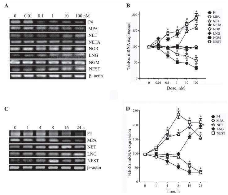
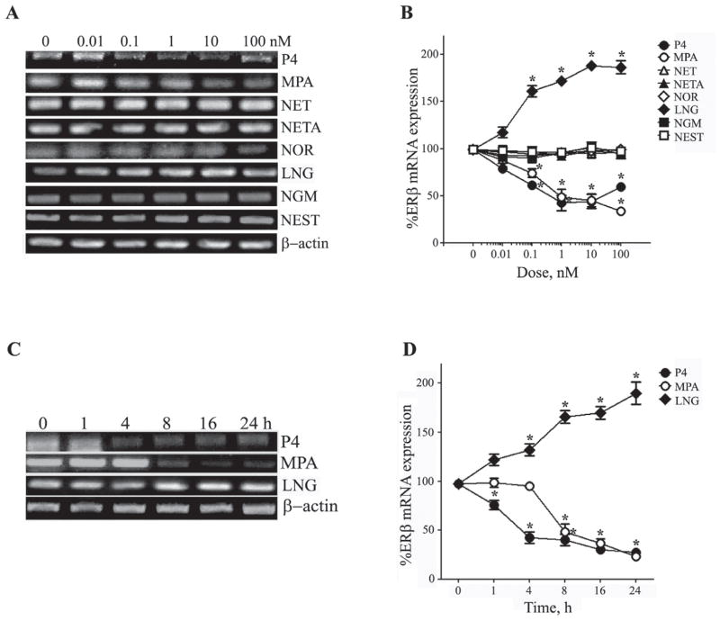
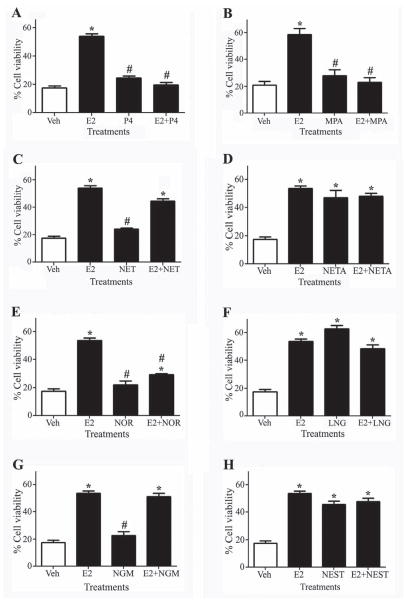
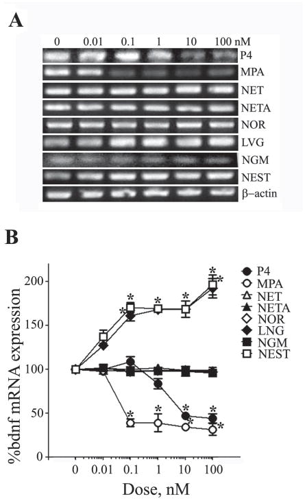
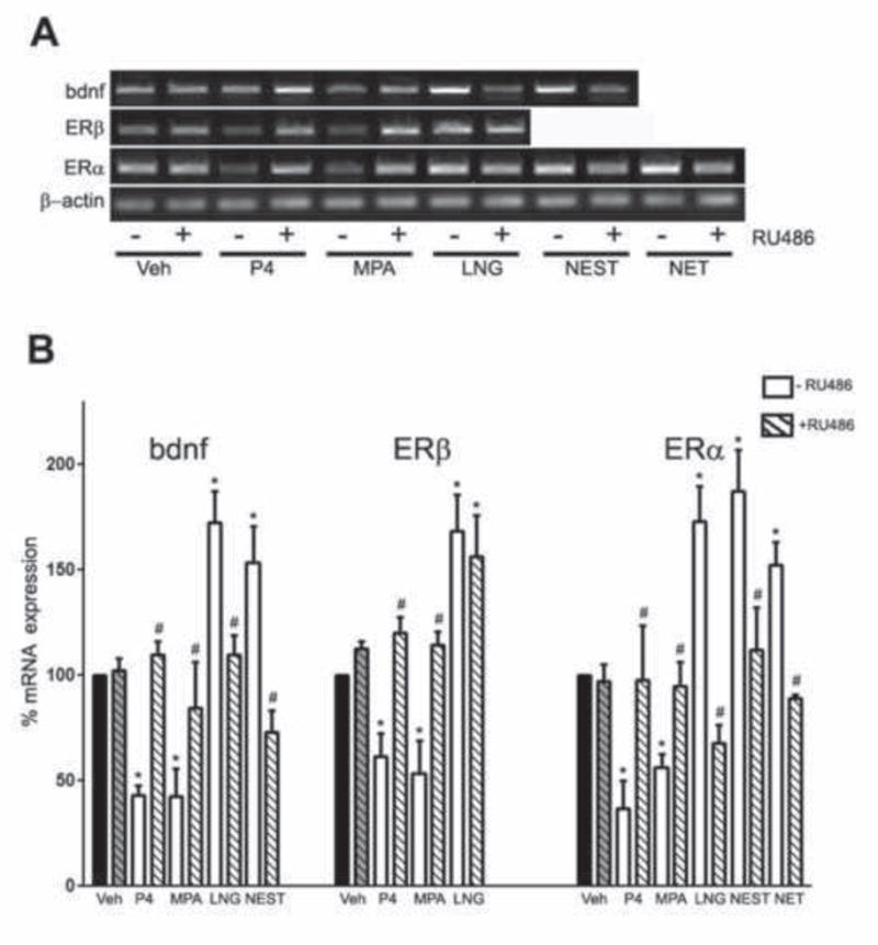
Progesterone and other progestagens are used in combination with estrogens for clinical purposes, including contraception and postmenopausal hormone therapy. Progesterone and estrogens have interactive effects in brain, however interactions between synthetic progestagens and 17β-estradiol (E2) in neurons are not well understood. In this study, we investigated the effects of seven clinically relevant progestagens on estrogen receptor (ER) mRNA expression, E2-induced neuroprotection, and E2-induced BDNF mRNA expression. We found that medroxyprogesterone acetate decreased both ERα and ERβ expression and blocked E2-mediated neuroprotection and BDNF expression. Conversely, levonorgestrel and nesterone increased ERα and or ERβ expression, were neuroprotective, and failed to attenuate E2-mediated increases in neuron survival and BDNF expression. Other progestagens tested, including norethindrone, norethindrone acetate, norethynodrel, and norgestimate, had variable effects on the measured endpoints. Our results demonstrate a range of qualitatively different actions of progestagens in cultured neurons, suggesting significant variability in the neural effects of clinically utilized progestagens.

Keywords: BDNF; Neuroprotection; Oestrogen receptor; Progestagen.


Figures

Figure 1
Figure 1. Progestagens regulate ERα mRNA expression in concentration and time-dependent manners
(A) Representative agarose gel of RT-PCR products qualitatively show relative changes in mRNA levels of the ERα induced by 24 h exposure to 0 – 100 nM of progestagens. β-actin was used as an internal control. (B) The relative levels of ERα after treatment with increasing progestagen concentrations were also determined quantitatively using real-time PCR. Data show the mean (±SEM) expression levels, relative to vehicle-treated controls after normalizing with corresponding values of β-actin. Representative agarose gel of RT-PCR (C) and quantitative graph from real-time PCR (D) shows changes in levels of ERα mRNA induced by 0 – 24 h exposure to 10 nM progestagens. Statistical significance is based on analysis of pooled raw data using ANOVA followed by the Fisher LSD. * P≤0.05 relative to corresponding vehicle-treated control group.
Figure 2
Figure 2. Progestagens regulate ERβ mRNA expression in concentration and time-dependent manners
(A) Representative agarose gel of RT-PCR products qualitatively show relative changes in mRNA levels of the ERβ induced by 24 h exposure to 0 – 100 nM of progestagens. β-actin was used as an internal control. (B) The relative levels of ERβ after treatment with increasing progestagen concentrations were also determined quantitatively using real-time PCR. Data show the mean (±SEM) expression levels, relative to vehicle-treated controls after normalizing with corresponding values of β-actin. Representative agarose gel of RT-PCR (C) and quantitative graph from real-time PCR (D) shows changes in levels of ERβ mRNA induced by 0 – 24 h exposure to 10 nM progestagens. Statistical significance is based on analysis of pooled raw data using ANOVA followed by the Fisher LSD. * P≤0.05 relative to corresponding vehicle-treated control group.
Figure 3
Figure 3. Progestagens regulate neuroprotection alone and in combination with E2
Neuronal cultures were treated with vehicle or 10 nM P4 (A), MPA (B), NET (C), NETA (D), NOR (E), LNG (F), NGM (G), and NEST (H) alone or in combination with 10 nM E2 for 2 h, followed by treatment with 3 μM Apoptosis Activator II (AAII) for 24 h, and processed for cell viability. Data show mean cell viability (± SEM) of a representative experiment (N = 3). * P≤0.05 relative to the vehicle-treated control condition exposed to AAII. # P≤0.05 relative to E2-treated condition exposed to AAII and is based on ANOVA followed by Fisher LSD.
Figure 4
Figure 4. Progestagens regulate BDNF mRNA expression in a concentration-dependent manner
(A) Representative agarose gel of RT-PCR products qualitatively show relative changes in mRNA levels of the BDNF induced by 24 h exposure to 0 – 100 nM of progestagens. β-actin was used as an internal control. (B) The relative levels of BDNF after treatment with increasing progestagen concentrations were also determined quantitatively using real-time PCR. Data show the mean (±SEM) expression levels, relative to vehicle-treated controls after normalizing with corresponding values of β-actin. * P≤0.05 relative to corresponding vehicle-treated condition.
Figure 5
Figure 5. Progestagens regulate E2-induced BDNF mRNA expression
(A) Representative agarose gel of RT-PCR products qualitatively shows changes in the levels of BDNF mRNA following treatment with vehicle, 10 nM E2, 10 nM progestagen, and 10 nM E2+progestagen. (B–I) Quantitative real-time PCR data show the mean (±SEM) expression levels compared to the vehicle-treated control group (white bar) for BDNF mRNA respectively. All data are normalized with corresponding β-actin values. Statistical significance is based on analysis of pooled raw data using ANOVA followed by the Fisher HSD. * P≤0.05 relative to the vehicle-treated control group. # P≤0.05 relative to the E2-treatment group.
Figure 6
Figure 6. Transcriptional regulation of progestagens is reversed by RU486
(A) Representative agarose gel of RT-PCR products qualitatively shows changes in the levels of BDNF, ERα, and ERβ mRNAs following treatment with 10 nM progestagen with or without 50 nM RU486. (B) Quantitative real-time PCR data show the mean (±SEM) expression levels compared to the vehicle-treated control group (black bar) for BDNF, ERα and ERβ mRNA respectively. All data are normalized with corresponding β-actin values. Statistical significance is based on analysis of pooled raw data using ANOVA followed by the Fisher HSD. * P≤0.05 relative to the vehicle-treated control group. # P≤0.05 relative to the corresponding –RU486 treatment group for a specific progestagen.

References

    1. Aguirre C, Jayaraman A, Pike C, Baudry M. Progesterone inhibits estrogen-mediated neuroprotection against excitotoxicity by down-regulating estrogen receptor-beta. J Neurochem. 2010;115(5):1277–87. - PMC - PubMed
    1. Aguirre CC, Baudry M. Progesterone reverses 17beta-estradiol-mediated neuroprotection and BDNF induction in cultured hippocampal slices. Eur J Neurosci. 2009;29(3):447–54. - PMC - PubMed
    1. Alkayed NJ, Murphy SJ, Traystman RJ, Hurn PD, Miller VM. Neuroprotective effects of female gonadal steroids in reproductively senescent female rats. Stroke. 2000;31(1):161–8. - PubMed
    1. Allen TK, Feng L, Grotegut CA, Murtha AP. Progesterone Receptor Membrane Component 1 as the Mediator of the Inhibitory Effect of Progestins on Cytokine-Induced Matrix Metalloproteinase 9 Activity In Vitro. Reprod Sci 2013 - PMC - PubMed
    1. Baudry M, Bi X, Aguirre C. Progesterone-estrogen interactions in synaptic plasticity and neuroprotection. Neuroscience. 2013;239:280–94. - PMC - PubMed

Publication types

MeSH terms

LinkOut - more resources