. 2014 Apr;23(4):423-32.
doi: 10.1002/pro.2424. Epub 2014 Mar 10.

Crystal structure of the transcriptional regulator Rv1219c of Mycobacterium tuberculosis

Affiliations

Crystal structure of the transcriptional regulator Rv1219c of Mycobacterium tuberculosis

Nitin Kumar et al. Protein Sci. 2014 Apr.

Abstract

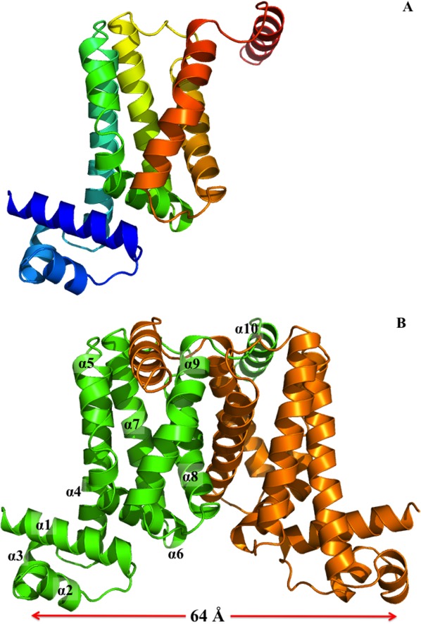
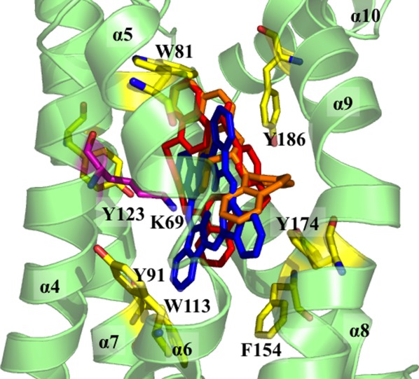
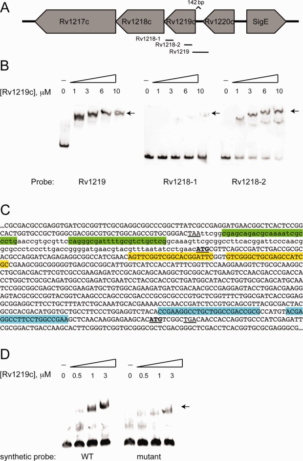
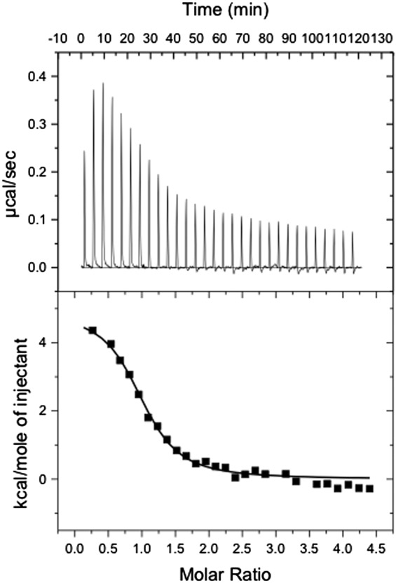
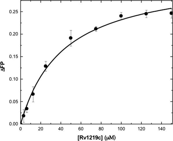
The Rv1217c-Rv1218c multidrug efflux system, which belongs to the ATP-binding cassette superfamily, recognizes and actively extrudes a variety of structurally unrelated toxic chemicals and mediates the intrinsic resistance to these antimicrobials in Mycobacterium tuberculosis. The expression of Rv1217c-Rv1218c is controlled by the TetR-like transcriptional regulator Rv1219c, which is encoded by a gene immediately upstream of rv1218c. To elucidate the structural basis of Rv1219c regulation, we have determined the crystal structure of Rv1219c, which reveals a dimeric two-domain molecule with an entirely helical architecture similar to members of the TetR family of transcriptional regulators. The N-terminal domains of the Rv1219c dimer are separated by a large center-to-center distance of 64 Å. The C-terminal domain of each protomer possesses a large cavity. Docking of small compounds to Rv1219c suggests that this large cavity forms a multidrug binding pocket, which can accommodate a variety of structurally unrelated antimicrobial agents. The internal wall of the multidrug binding site is surrounded by seven aromatic residues, indicating that drug binding may be governed by aromatic stacking interactions. In addition, fluorescence polarization reveals that Rv1219c binds drugs in the micromolar range.

Keywords: Mycobacterium tuberculosis; Rv1219c multidrug efflux regulator; multidrug resistance; transcriptional regulation.


Figures

Figure 1
Figure 1
Structure of the M. tuberculosis Rv1219c regulator. (A) Ribbon diagram of a protomer of Rv1219c. The molecule is colored using a rainbow gradient from the N-terminus (blue) to the C-terminus (red). An interactive view is available in the electronic version of the article. (B) Ribbon diagram of the Rv1219c dimer. Each subunit of Rv1219c is labeled with a different color (green and orange). The figure was prepared using PyMOL (http://www.pymol.sourceforge.net). An interactive view is available in the electronic version of the article.
Figure 2
Figure 2
The C-terminal multidrug binding site. The seven aromatic residues (W81, Y91, W113, Y123, F154, Y174, and Y186) that surround the interior of the multidrug binding cavity of Rv1219c are in yellow sticks. The cationic residue K69, which is thought to be important for interacting with the bound drug, is in magenta stick. The top three Rv1219c substrates are also included (red, UCL 1684; orange, (S)-(+)-N-(3,5-Dioxa-4-phosphacyclohepta[2,1-a;3,4-a′]dinaphthalen-4-yl)-dibenzo[b,f]azepine; blue, phthalocyanine).
Figure 3
Figure 3
Rv1219c binds to promoter regions of rv1219c and upstream of the rv1218c gene. (A) A schematic depicting the DNA probes used in electrophoretic mobility shift assays (EMSAs) to examine the promoter region of rv1219c and rv1218c. (B) EMSAs were performed using 12 nM Dig-labeled probe and the indicated micromolar concentrations of protein. An arrow denotes the shifted probes. (C) The reverse complement sequence of rv1219c-rv1218c region. The stop codon of rv1220c is underlined, the start codons of rv1219c and rv1218c are bold and underlined. The 142 bp intergenic region between rv1220c and rv1219c is in lowercase, coding sequences are in upper case. Indirect repeats were identified in each of the probes using the MEME algorithm. The high-affinity binding site in the rv1219c promoter probe is highlighted in green. The low-affinity Rv1219c binding motifs in the rv1218-1 and rv1218-2 probes are highlighted in blue and yellow, respectively. (D) EMSAs were performed using 12 nM Dig-labeled probe and the indicated micromolar concentrations of protein. An arrow denotes the shifted probes. Synthetic oligonucleotide duplexes comprising the high affinity binding site plus eight nucleotides on either side (74 bp total) were used as probes.
Figure 4
Figure 4
Representative isothermal titration calorimetry for the binding of the 58-bp DNA to Rv1219c regulator. (A) Each peak corresponds to the injection of 10 μL of 200 μM 58-bp DNA in buffer containing 10 mM Na-phosphate (pH 7.5) and 100 mM NaCl into the reaction cell containing 10 μM Rv1219c in the same buffer. (B) Cumulative heat of reaction is displayed as a function of the injection number. The solid line is the least-square fit to the experimental data, giving a KA of 1.2 ± 0.1 × 10⁻⁶ M−1. The molar-to-molar ratio of dimeric Rv1219c:58-bp ds-DNA is 1:1.
Figure 5
Figure 5
Representative fluorescence polarization of Rv1219c. The binding isotherm of Rv1219c with safranin O, showing a KD of 42.4 ± 7.6 μM. Fluorescence polarization is defined by the equation, FP = (VH)/(V + H), where FP equals polarization, V equals the vertical component of the emitted light, and H equals the horizontal component of the emitted light of a fluorophore when excited by vertical plane polarized light. FP is a dimensionless entity and is not dependent on the intensity of the emitted light or on the concentration of the fluorophore. mP is related to FP, where 1 mP equals one thousandth of a FP.

References

    1. Maartens G, Wilkinson RJ. Tuberculosis. Lancet. 2007;370:2030–2043. - PubMed
    1. World Health Organization. 2010. Fact sheet no. 104: tuberculosis. Available at http://www.who.int/mediacentre/factsheets/fs104/en/index.html.
    1. Centers for Disease Control and Prevention. 2012. Fact sheet: tuberculosis. Available at: http://www.cdc.gov/tb/topic/treatment/default.htm.
    1. Frieden TR, Sterling T, Pablos-Mendez A, Kilburn JO, Cauthen GM, Dooley SW. The emergence of drug-resistant tuberculosis in New York City. N Engl J Med. 1993;328:521–556. - PubMed
    1. Pillay M, Sturm AW. Evolution of the extensively drug-resistant F15/LAM4/KZN strain of Mycobacterium tuberculosis in KwaZulu-Natal, South Africa. Clin Infect Dis. 2007;45:1409–1414. - PubMed

Publication types

MeSH terms

Associated data

LinkOut - more resources