. 2014 Jan 28;111(4):1628-33.
doi: 10.1073/pnas.1315588111. Epub 2014 Jan 14.

Synthetic genetic array screen identifies PP2A as a therapeutic target in Mad2-overexpressing tumors

Affiliations

Synthetic genetic array screen identifies PP2A as a therapeutic target in Mad2-overexpressing tumors

Yang Bian et al. Proc Natl Acad Sci U S A. .

Abstract

The spindle checkpoint is essential to ensure proper chromosome segregation and thereby maintain genomic stability. Mitotic arrest deficiency 2 (Mad2), a critical component of the spindle checkpoint, is overexpressed in many cancer cells. Thus, we hypothesized that Mad2 overexpression could specifically make cancer cells susceptible to death by inducing a synthetic dosage lethality defect. Because the spindle checkpoint pathway is highly conserved between yeast and humans, we performed a synthetic genetic array analysis in yeast, which revealed that Mad2 overexpression induced lethality in 13 gene deletions. Among the human homologs of candidate genes, knockdown of PPP2R1A, a gene encoding a constant regulatory subunit of protein phosphatase 2, significantly inhibited the growth of Mad2-overexpressing tumor cells. PPP2R1A inhibition induced Mad2 phosphorylation and suppressed Mad2 protein levels. Depletion of PPP2R1A inhibited colony formation of Mad2-overexpressing HeLa cells but not of unphosphorylated Mad2 mutant-overexpressing cells, suggesting that the lethality induced by PP2A depletion in Mad2-overexpressing cells is dependent on Mad2 phosphorylation. Also, the PP2A inhibitor cantharidin induced Mad2 phosphorylation and inhibited the growth of Mad2-overexpressing cancer cells. Aurora B knockdown inhibited Mad2 phosphorylation in mitosis, resulting in the blocking of PPP2R1A inhibition-induced cell death. Taken together, our results strongly suggest that PP2A is a good therapeutic target in Mad2-overexpressing tumors.

Keywords: aneuploidy; anticancer drug; cancer therapy target; yeast genetics.


Conflict of interest statement

The authors declare no conflict of interest.

Figures
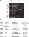
Fig. 1.
Fig. 1.
Synthetic genetic array screen identifies candidate genes whose deletion kills MAD2-overexpressing yeast cells. (A) The yeast haploid deletion MATa library (4,541 strains) screen identified 18 candidate genes. The 18 deletion mutant strains carrying pGAL-MAD2 were spotted onto dextrose (DEX; negative control) or galactose plates (GAL). The deletion mutant strains carrying pGAL only were spotted onto galactose plates as a negative control. (B) The list of yeast candidate genes whose deletion causes synthetic dosage lethality and their human homologs. The E-values were obtained by Blast searches by using BIOBASE’s biological databases and the NCBI Blast search.
Fig. 2.
Fig. 2.
Synthetic lethality with Mad2 overexpression in HeLa cells. (A) Cell lysates from HeLa or Mad2-overexpressing HeLa (HeLa-Mad2 O/E) cells were immunoblotted with anti-Mad2 antibody or Hsc70 antibody (a loading control). (B) A schematic figure of the assay. GFP-expressing HeLa cells and Mad2-overexpressing HeLa cells were mixed in a 1:1 ratio. Mixed cells were transfected with the candidate gene’s siRNA. After 72 h, the ratio of Mad2-overexpressing cells to HeLa-GFP cells was measured by FACS. (C) The percentages of Mad2-overexpressing cells were calculated over GFP-expressing cells. The relative percentages of Mad2-overexpressing cells were normalized against luciferase siRNA-treated cells.
Fig. 3.
Fig. 3.
PPP2R1A depletion inhibits colony formation of Mad2-overexpressing cells. (A) HeLa or Mad2-overexpressing HeLa cells (HeLa-Mad2 O/E) were transfected with luciferase siRNA (Luc) or PPP2R1A siRNA. After 3 d, 2,000 cells were split per well of a six-well plate, and cell images were taken after 10 d. (B) The colony numbers were counted in A. (C) hTERT-RPE1 or Mad2-overexpressing hTERT-RPE1 cells were transfected with luciferase siRNA (Luc) or PPP2R1A siRNA. After 3 d, 1,000 cells were split per well of a six-well plate, and cell images were taken after 10 d. (D) The colony numbers were counted in C. (E) hTERT-RPE1 or Mad2-overexpressing hTERT-RPE1 cell lysates were immunoblotted with anti-Mad2 antibody or anti-GAPDH antibody. (F) HeLa cells were transfected with luciferase siRNA (Luc) or PPP2R1A siRNA and immunoblotted with anti-PPP2R1A antibody or anti-GAPDH antibody.
Fig. 4.
Fig. 4.
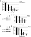
Cantharidin inhibits the viability of Mad2-overexpressing tumor cells. (A) Mad2-overexpressing HeLa cells or HeLa cells were split to 800 cells per well of a six-well plate, and cantharidin was added at indicated concentrations the next day. Colony numbers were counted after ∼10 d. (B) HeLa cells or OS-17 cell lysates were immunoblotted with anti-Mad2 antibody or anti-Hsc70 antibody. (C) HeLa cells or OS-17 cells were split to 1,000 cells per well of a six-well plate, and cantharidin was added at indicated concentrations the next day. Colony numbers were counted after colony formation. (D) HeLa cells or Weri1 cell lysates were immunoblotted with anti-Mad2 antibody or Hsc70 antibody. (E) Cells were split into a 96-well plate after cantharidin treatment, and cell viability was assessed by the MTT assay.
Fig. 5.
Fig. 5.
The mitotic arrest caused by PP2A inhibition requires an unperturbed spindle checkpoint. (A) HeLa or Mad2-overexpressing HeLa cells (HeLa-Mad2 O/E) were spread on coverslips. The next day, cells were transfected with luciferase siRNA or PPP2R1A siRNA. After 3 d, cells were stained with mouse monoclonal anti-phosphorylated histone H3 (p-H3) antibodies and FITC-conjugated secondary antibodies (green). DNA was stained with DAPI (blue) to visualize prophase, prometaphase, and metaphase cells. Tubulin was stained with mouse anti-β tubulin antibody and Alexa 594–conjugated secondary antibodies (red) to further confirm the cell cycle stages. (B) The number of mitotic cells was counted among more than 600 cells. Mitotic cells were p-H3+ and had a characteristic chromosome morphology. (C) HeLa cells were transfected with control luciferase siRNA or BubR1 siRNA. After 48 h, cells were treated with DMSO, nocodazole (250 ng/mL), or cantharidin (2.5 μM) for 16 h. Cells were stained with mouse p-H3 antibody and FITC-conjugated secondary antibodies (green). The number of mitotic cells was counted by fluorescence microscopy. (D) HeLa cell lysates were immunoprecipitated with anti-Mad2, anti-PPP2R1A, or IgG control antibodies and immunoblotted to analyze endogenous associated proteins.
Fig. 6.
Fig. 6.
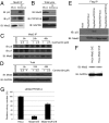
PP2A dephosphorylates Mad2. (A) HeLa cells were transfected with luciferase siRNA or PPP2R1A siRNA. After 3 d, cell lysates were immunoprecipitated with anti-Mad2 antibody, and immunoblotted with anti-phosphoserine antibody or anti-phosphothreonine antibody. (B) Total lysates for A were immunoblotted with anti-Mad2 antibody, anti-GAPDH (control), or anti-PPP2R1A antibody. (C) HeLa cells were treated with cantharidin at indicated concentrations and collected at 8, 24, or 48 h. Cell lysates were immunoprecipitated with anti-Mad2 antibody and immunoblotted with the same antibody as in A. (D) Total lysates for C were immunoblotted with anti-Mad2 antibody or anti-GAPDH. (E) HeLa cells were transfected with Flag-Mad2–expressing plasmid or vector and transfected with indicated siRNA. After 48 h, lysates were immunoprecipitated by ANTI-FLAG M2 Affinity Gel, and the immunoprecipitates were blotted with anti-phosphoserine antibody or anti-flag antibody. (F) HeLa or Mad2-4SA–overexpressing HeLa cell lysates were immunoblotted with anti-Mad2 antibody or anti-GAPDH antibody. (G) HeLa cells, Mad2-overexpressing HeLa (Mad2 O/E) cells, or Mad2-4SA cells were transfected with luciferase siRNA or PPP2R1A siRNA. After 3 d, cells were split to 2,000 cells per well, and colony numbers were counted after ∼10 d.
Fig. 7.
Fig. 7.
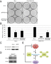
Mad2 phosphorylation pathways. (A) HeLa cells or Mad2-overexpressing (HeLa Mad2 O/E) cells were transfected with luciferase siRNA or PPP2R1A siRNA with or without Aurora B siRNA. After 2 d, 2,000 cells were split per well of a six-well plate, and colony images were taken after 10 d. (B) The colony numbers in A were counted. (C) HeLa cells were transfected with luciferase siRNA or Aurora B siRNA. After 3 d, cell lysates were immunoblotted with Aurora B antibody or GAPDH antibody. (D) HeLa cells were transfected with luciferase siRNA or Aurora B siRNA. After 2 d, cells were treated with nocodazole, and cell lysates were immunoprecipitated with anti-Mad2 antibody and immunoblotted with a phosphoserine antibody after 8 h. (E) Model of Mad2 phosphorylation.

References

    1. Li R, Murray AW. Feedback control of mitosis in budding yeast. Cell. 1991;66(3):519–531. - PubMed
    1. Li Y, Benezra R. Identification of a human mitotic checkpoint gene: hsMAD2. Science. 1996;274(5285):246–248. - PubMed
    1. Cahill DP, et al. Mutations of mitotic checkpoint genes in human cancers. Nature. 1998;392(6673):300–303. - PubMed
    1. Sotillo R, et al. Mad2 overexpression promotes aneuploidy and tumorigenesis in mice. Cancer Cell. 2007;11(1):9–23. - PMC - PubMed
    1. Schvartzman JM, Sotillo R, Benezra R. Mitotic chromosomal instability and cancer: Mouse modelling of the human disease. Nat Rev Cancer. 2010;10(2):102–115. - PMC - PubMed

Publication types