. 2014 Feb 4;55(2):737-44.
doi: 10.1167/iovs.13-13429.

Quantitative analyses of retinal vascular area and density after different methods to reduce VEGF in a rat model of retinopathy of prematurity

Affiliations

Quantitative analyses of retinal vascular area and density after different methods to reduce VEGF in a rat model of retinopathy of prematurity

Haibo Wang et al. Invest Ophthalmol Vis Sci. .

Abstract

Purpose: Targeted inhibition of Müller cell (MC)-produced VEGF or broad inhibition of VEGF with an intravitreal anti-VEGF antibody reduces intravitreal neovascularization in a rat model of retinopathy of prematurity (ROP). In this study, we compared the effects of these two approaches on retinal vascular development and capillary density in the inner and deep plexi in the rat ROP model.

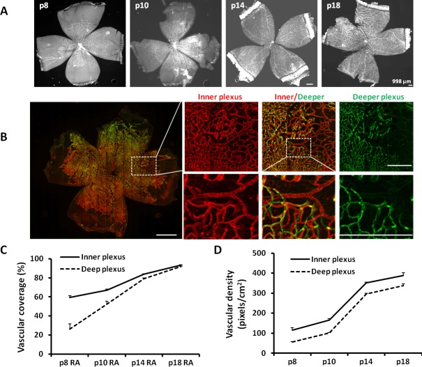
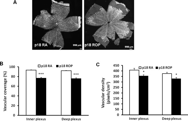
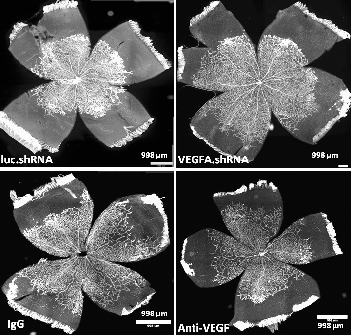
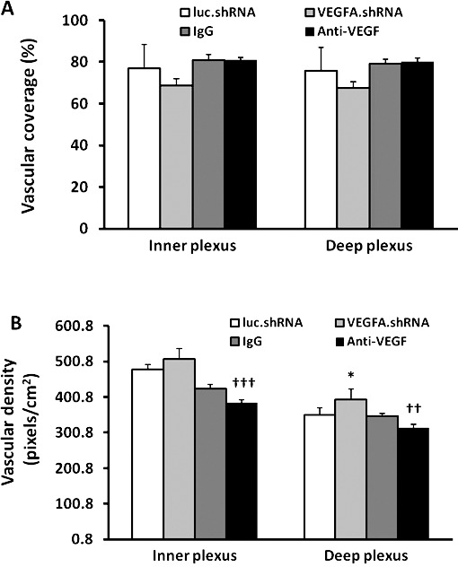
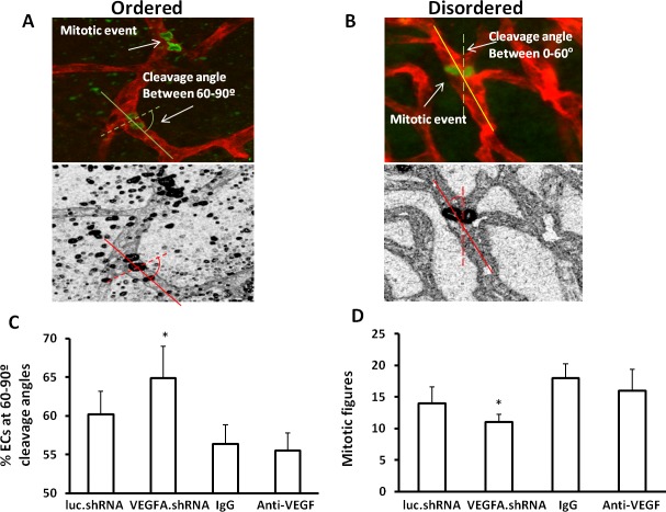
Methods: In the rat model of ROP, pups received 1 μL of (1) subretinal lentivector-driven short hairpin RNA (shRNA) to knockdown MC-VEGFA (VEGFA.shRNA) or control luciferase shRNA, or (2) intravitreal anti-VEGF antibody (anti-VEGF) or control isotype goat immunoglobulin G (IgG). Analyses of lectin-stained flat mounts at postnatal day 18 (p18) included: vascular/total retinal areas (retinal vascular coverage) and pixels of fluorescence/total retinal area (capillary density) of the inner and deep plexi determined with the Syncroscan microscope, and angles between cleavage planes of mitotic vascular figures labeled with anti-phosphohistone H3 and vessel length.

Results: Retinal vascular coverage and density increased in both plexi between p8 and p18 in room air (RA) pups. Compared with RA, p18 ROP pups had reduced vascular coverage and density of both plexi. Compared with respective controls, VEGFA.shRNA treatment significantly increased vascular density in the deep plexus, whereas anti-VEGF reduced vascular density in the inner and deep plexi. Vascular endothelial growth factor-A.shRNA caused more cleavage angles predicting vessel elongation and fewer mitotic figures, whereas anti-VEGF treatment led to patterns of pathologic angiogenesis.

Conclusions: Targeted treatment with lentivector-driven VEGFA.shRNA permitted physiologic vascularization of the vascular plexi and restored normal orientation of dividing vascular cells, suggesting that regulation of VEGF signaling by targeted treatment may be beneficial.

Keywords: VEGF; intrvitreal neovscularization; rat model of retinopathy of prematurity; vascular density; vascular extent.


Figures

Figure 1
Figure 1
Retinal vascular coverage and density in the inner and deeper plexi are increased during development at p8, p10, p14, and p18 in RA. (A) Syncroscan images of lectin-stained retinal flat mounts. (B) A portion of p18 VEGFA.shRNA-treated flat mount in the ROP model showing inner plexus, deep plexus, and combined image of both plexi assigned different colors (inner-red, deep-green; inner and deep plexi offset to permit visualization; lines indicate magnification size at the same unit). (C) Vascular coverage determined by vascular area normalized to total retina area. (D) Vascular density determined by the number of pixels of lectin fluorescence normalized to total retinal area (overall one-way ANOVA ***P < 0.001; results are means ± SD).
Figure 2
Figure 2
Retinal vascular coverage and density are reduced in both inner and deep plexi of pups raised in the ROP model at p18. (A) Images of retinal flat mounts of pups raised in RA or exposed to the ROP model. (B) Retinal vascular coverage and (C) retinal vascular density in the inner and deep plexi (*P < 0.05, ***P < 0.001 versus RA, two-way ANOVA; results are means ± SD).
Figure 3
Figure 3
Vascular endothelial growth factor-A.shRNA versus anti-VEGF treatments on retinal vascular morphology. Images of lectin-stained retinal flat mounts from pups treated with luc.shRNA, VEGFA.shRNA, IgG, and anti-VEGF at p18 in the ROP model.
Figure 4
Figure 4
Retinal vascular coverage and density in the inner and deep plexi of pups treated with VEGFA.shRNA or anti-VEGF in the ROP model at p18 compared with respective controls. (A) Retinal vascular coverage and (B) vascular density (*P < 0.05 versus luc.shRNA; ††P < 0.01 and †††P < 0.001 versus IgG, two-way ANOVA; results are means ± SD).
Figure 5
Figure 5
Vascular endothelial growth factor-A.shRNA treatment reduces disordered angiogenesis in the ROP model. Diagram of cleavage angles produced from lectin-stained flat mounts colabeled for phosphohistone H3 showing cells in anaphase: (A) cleavage angles between 60° and 90° predict ordered angiogenesis; (B) cleavage angles between 0° and 60° predict widened or disordered angiogenesis; (C) percentage of vascular cells with cleavage angles between 60° and 90°; and (D) total number of mitotic figures determined as phosphohistone H3–labeled vascular cells in retinal flat mounts from pups treated with luc.shRNA, VEGFA.shRNA, IgG, or anti-VEGF (*P < 0.05 versus luc.shRNA, two-way ANOVA; results are means ± SD).

References

    1. Chan-Ling T, Gock B, Stone J. Supplemental oxygen therapy: basis for noninvasive treatment of retinopathy of prematurity. Invest Ophthalmol Vis Sci. 1995; 36: 1215–1230 - PubMed
    1. Aiello LP, Avery RL, Arrigg PG, et al. Vascular endothelial growth factor in ocular fluid of patients with diabetic retinopathy and other retinal disorders. N Engl J Med. 1994; 331: 1480–1487 - PubMed
    1. Gardiner TA, Gibson DS, de Gooyer TE, de la Cruz VF, McDonald DM, Stitt AW. Inhibition of tumor necrosis factor-{alpha} improves physiological angiogenesis and reduces pathological neovascularization in ischemic retinopathy. Am J Pathol. 2005; 166: 637–644 - PMC - PubMed
    1. Saito Y, Geisen P, Uppal A, Hartnett ME. Inhibition of NAD(P)H oxidase reduces apoptosis and avascular retina in an animal model of retinopathy of prematurity. Mol Vis. 2007; 13: 840–853 - PMC - PubMed
    1. Ema M, Taya S, Yokotani N, Matsuda Y, Fujii-Kuriyama Y. A novel bHLH-PAS factor with close sequence similarity to hypoxia inducible factor 1 alpha regulates VEGF expression and is potentially involved in lung and vascular development. Proc Natl Acad Sci U S A. 1997; 94: 4273–4278 - PMC - PubMed

Publication types

MeSH terms

Substances