. 2014 Mar 14;289(11):7307-19.
doi: 10.1074/jbc.M113.537878. Epub 2014 Jan 14.

CD2-associated protein (CD2AP) enhances casitas B lineage lymphoma-3/c (Cbl-3/c)-mediated Ret isoform-specific ubiquitination and degradation via its amino-terminal Src homology 3 domains

Affiliations

CD2-associated protein (CD2AP) enhances casitas B lineage lymphoma-3/c (Cbl-3/c)-mediated Ret isoform-specific ubiquitination and degradation via its amino-terminal Src homology 3 domains

Gina N Calco et al. J Biol Chem. .

Abstract

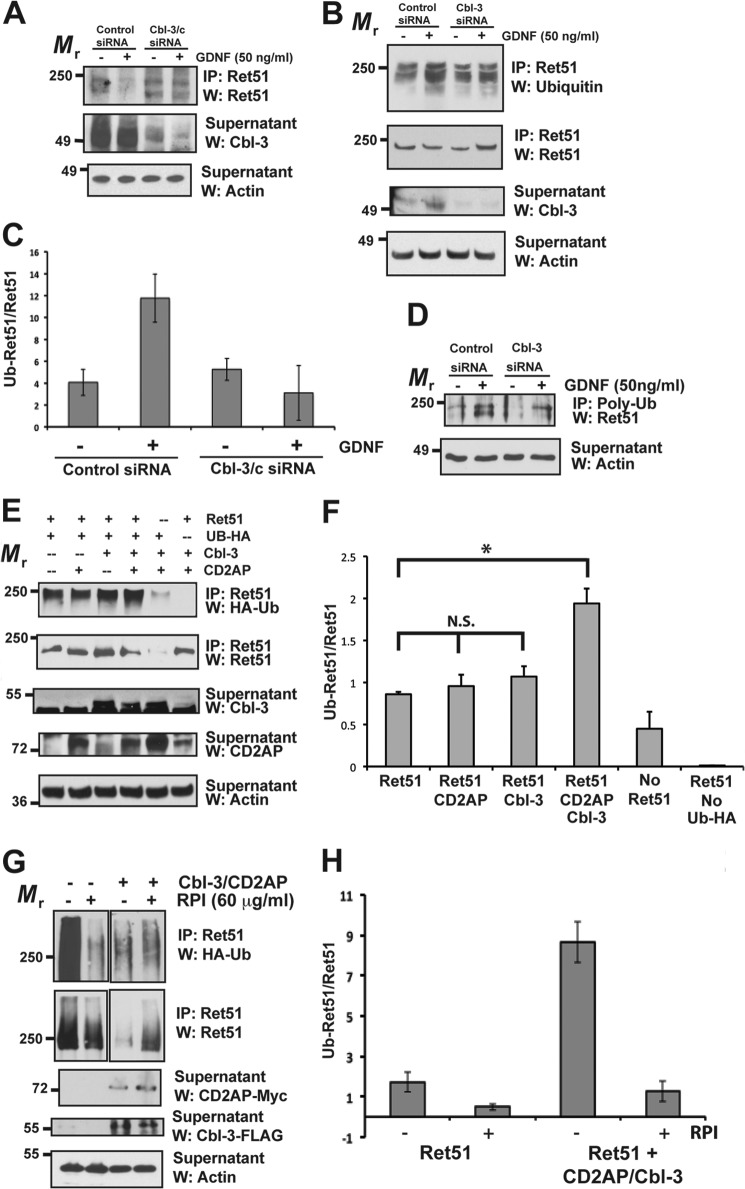
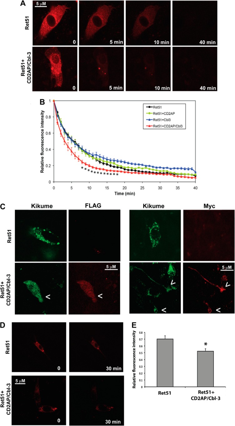
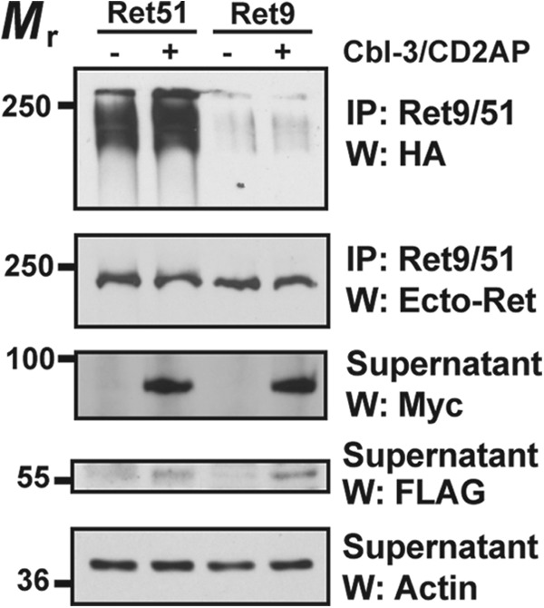
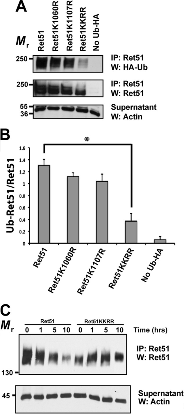
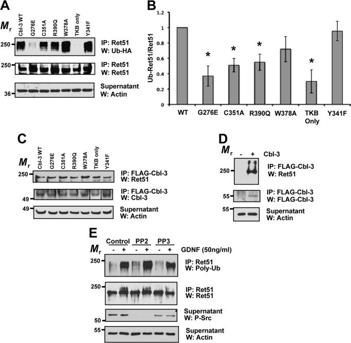
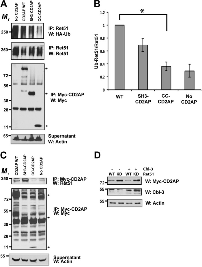
Ret is the receptor tyrosine kinase for the glial cell line-derived neurotrophic factor (GDNF) family of neuronal growth factors. Upon activation by GDNF, Ret is rapidly polyubiquitinated and degraded. This degradation process is isoform-selective, with the longer Ret51 isoform exhibiting different degradation kinetics than the shorter isoform, Ret9. In sympathetic neurons, Ret degradation is induced, at least in part, by a complex consisting of the adaptor protein CD2AP and the E3-ligase Cbl-3/c. Knockdown of Cbl-3/c using siRNA reduced the GDNF-induced ubiquitination and degradation of Ret51 in neurons and podocytes, suggesting that Cbl-3/c was a predominant E3 ligase for Ret. Coexpression of CD2AP with Cbl-3/c augmented the ubiquitination of Ret51 as compared with the expression of Cbl-3/c alone. Ret51 ubiquitination by the CD2AP·Cbl-3/c complex required a functional ring finger and TKB domain in Cbl-3/c. The SH3 domains of CD2AP were sufficient to drive the Cbl-3/c-dependent ubiquitination of Ret51, whereas the carboxyl-terminal coiled-coil domain of CD2AP was dispensable. Interestingly, activated Ret induced the degradation of CD2AP, but not Cbl-3/c, suggesting a potential inhibitory feedback mechanism. There were only two major ubiquitination sites in Ret51, Lys(1060) and Lys(1107), and the combined mutation of these lysines almost completely eliminated both the ubiquitination and degradation of Ret51. Ret9 was not ubiquitinated by the CD2AP·Cbl-3/c complex, suggesting that Ret9 was down-regulated by a fundamentally different mechanism. Taken together, these results suggest that only the SH3 domains of CD2AP were necessary to enhance the E3 ligase activity of Cbl-3/c toward Ret51.

Keywords: Neurodevelopment; Neurotrophic Factor; Protein Degradation; Receptor Tyrosine Kinase; Ubiquitination.


Figures

FIGURE 1.
FIGURE 1.
Cbl-3 and CD2AP together promote the ubiquitination of Ret51. A, differentiated mouse podocytes were transfected with siRNA to either Cbl-3/c, or a scrambled, nonsilencing control siRNA (indicated above blots). After 48 h the cells were then stimulated with GDNF, or medium alone, for 24 h. Ret51 was immunoprecipitated from detergent extracts of cells subjected to these treatments, and the level of Ret degradation was determined by immunoblotting these immunoprecipitates with Ret51 antibodies (top panel). Cbl-3 immunoblotting (middle panel) of the supernatants displayed a reduction in Cbl-3 protein upon transfection with Cbl-3 siRNA. Actin immunoblotting of the supernatants confirmed that similar amounts of protein extracts were analyzed (bottom panel). B, primary sympathetic neurons were subjected to siRNA silencing of Cbl-3/c, as was done in A, and were stimulated with either GDNF or medium alone for 10 min, prior to any significant reduction in Ret51 levels. The level of Ret51 ubiquitination was determined by immunoblotting Ret51 immunoprecipitates derived from these neurons with antibodies to multiubiquitin (top panel). The amount of Ret51 in each condition was determined by reprobing the Ret51 immunoprecipitates with Ret 51 antibodies (upper middle panel). Cbl-3 immunoblotting of the supernatants from the immunoprecipitations confirmed that the transfection of siRNA targeted to Cbl-3/c reduced Cbl-3/c levels (lower middle panel), and actin immunoblotting of the supernatants again confirmed the analysis of equal amounts of protein (bottom panel). C, duplicate experiments were quantified and graphed as the means ± range. D, sympathetic neurons were subjected to Cbl-3 silencing, as in A, and ubiquitinated proteins were immunoprecipitated using polyubiquitin antibodies and probed with Ret51 antibodies. Actin confirmed the analysis of equal amounts of protein. E, NIH3T3 cells were transfected with CD2AP, Cbl-3/c, or CD2AP + Cbl-3/c along with Ret51 and HA-tagged ubiquitin. After 48 h, the cells were treated with proteasome and lysosome inhibitors and then detergent-extracted after an additional 6–12 h. The level of Ret51 ubiquitination in each condition was ascertained by immunoblotting Ret51 immunoprecipitates derived from these transfected cells with HA epitope tag antibodies (top panel). The level of Ret51, along with the levels of Cbl-3/c and CD2AP, were determined by immunoblotting, as was done in the middle panels of B. Actin immunoblotting of the supernatants served as a confirmation that similar amounts of protein were analyzed (bottom panel). F, quantifications of this experiment are shown as the means ± S.E. (three experiments were quantified). The asterisk indicates a statistically significant difference between the two conditions (p < 0.05), and N.S. indicates no significant difference. G, NIH3T3 cells were transfected with CD2AP and Cbl-3/c, or neither, along with Ret51 and HA-tagged ubiquitin. After 48 h, the cells were treated with proteasome and lysosome inhibitors along with the Ret kinase inhibitor RPI-1 (60 μm) and then detergent-extracted after an additional 6–12 h. H, Ret ubiquitination was monitored as in E, and the results were quantified. The data are presented as the means ± range. The experiments in this figure were repeated two or three times with similar results. IP, immunoprecipitation; W, Western blot.
FIGURE 2.
FIGURE 2.
CD2AP and Cbl-3/c alter Ret51 degradation in living cells. NIH3T3 cells were transfected with a Ret51-kikume fusion protein, and the presence of this protein was monitored by live cell, resonance scanning confocal microscopy. At t = 0, the kikume protein was photoconverted from green to red by exposure to 380-nm light, and z-stack images were taken every 15 s. In A, a typical time course of the loss of Ret51-kikume fluorescence from the cell is shown in cells transfected with Ret51-kikume alone and Ret51-kikume along with CD2AP and Cbl-3/c. This imaging method was applied to cells expressing Ret51-kikume alone, Ret51-kikume along with CD2AP, Cbl-3/c, or both CD2AP and Cbl-3/c. B, relative fluorescence intensity was measured from 12–15 cells from three to five independent transfections and graphed as the means ± S.E. (data points were only shown for each minute). Cells expressing CD2AP and Cbl-3/c (red triangles) displayed a significant increase in the rate of Ret51 degradation. Asterisks below the trace indicate a statistically significant difference (p < 0.05) as compared with Ret alone (black circles). Cells expressing Cbl-3/c (blue triangles) had a significantly slower rate of Ret51-kikume degradation, and expression of CD2AP was not significantly different from Ret51-kikume alone (green and black circles, respectively). C, to confirm the coexpresssion of all three proteins in these experiments, NIH3T3 cells were transfected with Ret51-kikume or Ret51-kikume along with CD2AP (Myc-tagged) and Cbl-3 (FLAG-tagged) and then fixed and immunolabeled with antibodies to Myc or FLAG (red). Because anti-Myc and anti-FLAG were both mouse monoclonal antibodies, triple labeling could not be performed. D, the cells were transfected with either Ret51-kikume alone or Ret51-kikume with CD2AP and Cbl-3. At t = 0, the cells were photoconverted but only imaged twice, once immediately following photoconversion and once after 30 min. E, the data from multiple cells of each condition were quantified. The relative fluorescence of Ret51-kikume was significantly lower (p < 0.05) in the presence of CD2AP and Cbl-3.
FIGURE 3.
FIGURE 3.
CD2AP and Cbl-3 are not regulators of Ret9 ubiquitination. NIH3T3 cells were transfected with either Ret51 or Ret9, along CD2AP and Cbl-3/c (indicated as +) or with neither (indicated as −). All conditions were also transfected with HA-ubiquitin. The extent of Ret9 and Ret51 ubiquitination was ascertained as in Fig. 1E (top panel). Immunoblotting with antibodies to the shared extracellular domain of Ret was performed on the immunoprecipitates to confirm that similar amounts of Ret51 and Ret9 were expressed (middle panel). Proteasome and lysosome inhibitors were applied to the cells 6–12 h before detergent extraction to stabilize the amount of Ret in each condition, as was done in Fig. 1E. Actin Westerns of the supernatants indicated that there were equal amounts of protein in each sample (bottom panel). The expression of CD2AP and Cbl-3/c were confirmed by Myc and FLAG immunoblotting, respectively, of the supernatants. This experiment was repeated four times with similar results. IP, immunoprecipitation; W, Western blot.
FIGURE 4.
FIGURE 4.
The major ubiquitination sites in Ret51 are lysines 1060 and 1107. A, NIH3T3 cells were transfected with CD2AP, Cbl-3, HA-ubiquitin, and either Ret51, Ret51-K1060R, Ret51-K1107R, or Ret51-K1060R/K1107R (indicated as KKRR). The amount of Ret51 ubiquitination was then ascertained as in Fig. 1E, including the use of proteasome and lysosome inhibitors to stop Ret51 degradation. Ret51 reprobing of the immunoprecipitates (middle panel), along with actin immunoblotting of the supernatants (bottom panel), were used to confirm that a similar amount of Ret51 and total protein were analyzed in each condition. B, the results in A were quantified as in Fig. 1F. The asterisk indicates a significant difference, p < 0.05. C, NIH3T3 cells were transfected with either Ret51-KKRR or Ret51, along with CD2AP and Cbl-3. After 48 h, the cells were given cycloheximide (1 μm) for 1, 5, or 10 h. The level of Ret51 in each condition was ascertained by immunoprecipitation of Ret51 followed by immunoblotting with Ret51 antibodies. Actin Westerns of the supernatants again confirmed the analysis of similar amounts of protein. Cycloheximide treatment inhibits protein synthesis and any further Ret51 production, thereby allowing the rate at which Ret51 is degraded to be determined. These experiments were repeated three times with similar results. IP, immunoprecipitation; W, Western blot.
FIGURE 5.
FIGURE 5.
Cbl-3/c-mediated ubiquitination of Ret51 requires both its ring finger and TKB domains. Cbl-3/c mutants were transfected into NIH3T3 cells along with CD2AP, Ret51, and HA-ubiquitin. These inactivating mutations included three point mutations in the ring finger domain of the E3 ligase (C351A, R390Q, and W378A) as well as a deletion of the entire ring finger (TKB only), a mutation of the phosphotyrosine-binding domain of the TKB region (G276E) and a Y341F mutant that eliminates a phosphorylation site that is conserved among Cbl family members. Wild type Cbl-3/c was also included as a positive control (each variant is indicated above the top panel). A, the extent of Ret51 ubiquitination was evaluated as was done in Fig. 1E and displayed. B, three representative experiments were quantified and graphed as the means ± S.E. Asterisks indicated a statistically significant difference (p < 0.05) between the indicated condition and wild type Cbl-3 (WT). C, identical transfections and inhibitor treatments were performed as in A. The cell extracts were subjected to FLAG immunoprecipitation to isolate the transfected Cbl-3 proteins that were all FLAG-tagged. These immunoprecipitations were then immunoblotted with Ret51 antibodies followed by Cbl-3 antibodies. Although the TKB domain of Cbl-3 has a smaller relative molecular mass, it is displayed in the panel along with the other Cbl-3/c mutants (box). Actin was used as a loading control. D, as a control for the specificity of the immunoprecipitations, cells were transfected with Ret and CD2AP in the presence or absence of FLAG-Cbl-3/c. FLAG immunoprecipitations and subsequent immunoblot analysis were performed as in C. E, primary sympathetic neurons were exposed to PP2, PP3, or vehicle alone (DMSO) for 1 h prior to GDNF stimulation for 10 min (indicated above blots). Ret51 was immunoprecipitated and then analyzed with polyubiquitin immunoblotting, as was done in Fig. 1B. Phospho-Src immunoblotting of the supernatants (third panel) confirmed inhibition of Src with PP2. The experiments shown in this figure were performed two or three times with similar results. IP, immunoprecipitation; W, Western blot.
FIGURE 6.
FIGURE 6.
The SH3 domains of CD2AP are sufficient for Ret association and Cbl-3-mediated ubiquitination. A, cells were transfected with full-length CD2AP, with the amino-terminal three SH3 domains of CD2AP (1–328 amino acids, SH3-CD2AP) or the carboxyl-terminal 120 amino acids that encodes the coiled-coil domain of CD2AP (561–681, CC-CD2AP). All of the cells were also cotransfected with Cbl-3, Ret51, and HA-ubiquitin. Ret ubiquitination was ascertained as in Fig. 1E. Supernatants from the first Ret immunoprecipitation were subsequently immunoprecipitated using anti-Myc and probed for Myc to confirm similar expression of the CD2AP variants. Asterisks on the right side of the Myc-CD2AP blot indicate the position of full-length CD2AP and the two truncations of CD2AP. B, the experiments in A were quantified and graphed as the means ± S.E. (n = 4). The asterisk indicates a significant difference between the WT CD2AP and CC-CD2AP conditions, p < 0.05. C, NIH3T3 cells were transfected with the same plasmids as in A. The cells were then subjected to Myc (CD2AP) immunoprecipitation followed by immunoblotting for Ret51. Myc immunoblotting of the immunoprecipitation supernatants confirmed the expression of the CD2AP variants (middle panels). The relative molecular masses of CD2AP, SH3-CD2AP, and CC-CD2AP are indicated with asterisks as in A (79–90, 45–55, and 10–15 kDa, respectively). Actin served as a loading control. D, NIH3T3 cells were transfected with Ret51 (WT) or kinase inactive Ret51 (KD) along with CD2AP, with or without Cbl-3. After 24 h, detergent extracts were produced from the transfected cells, and these extracts were immunoblotted with antibodies to CD2AP (Myc), Cbl-3, or actin as a loading control. The cells were not treated with protease inhibitors such that differences in protein degradation could be observed. The experiments in this figure were performed two or three times with similar results. IP, immunoprecipitation; W, Western blot.

Similar articles

Cited by

References

    1. Airaksinen M. S., Saarma M. (2002) The GDNF family. Signalling, biological functions and therapeutic value. Nat. Rev. Neurosci. 3, 383–394 - PubMed
    1. Baloh R. H., Enomoto H., Johnson E. M., Jr., Milbrandt J. (2000) The GDNF family ligands and receptors-implications for neural development. Curr. Opin. Neurobiol. 10, 103–110 - PubMed
    1. Bespalov M. M., Saarma M. (2007) GDNF family receptor complexes are emerging drug targets. Trends Pharmacol. Sci. 28, 68–74 - PubMed
    1. Wells S. A., Jr., Santoro M. (2009) Targeting the RET pathway in thyroid cancer. Clin. Cancer Res. 15, 7119–7123 - PubMed
    1. Pierchala B. A., Milbrandt J., Johnson E. M., Jr. (2006) Glial cell line-derived neurotrophic factor-dependent recruitment of Ret into lipid rafts enhances signaling by partitioning Ret from proteasome-dependent degradation. J. Neurosci. 26, 2777–2787 - PMC - PubMed

Publication types

MeSH terms