Review
. 2013 Dec 31:4:305.
doi: 10.3389/fgene.2013.00305.

Stress-induced ECM alteration modulates cellular microRNAs that feedback to readjust the extracellular environment and cell behavior

Affiliations
Review

Stress-induced ECM alteration modulates cellular microRNAs that feedback to readjust the extracellular environment and cell behavior

Evgeniia V Edeleva et al. Front Genet. .

Abstract

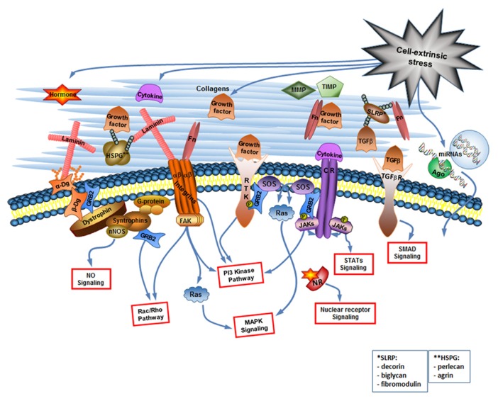
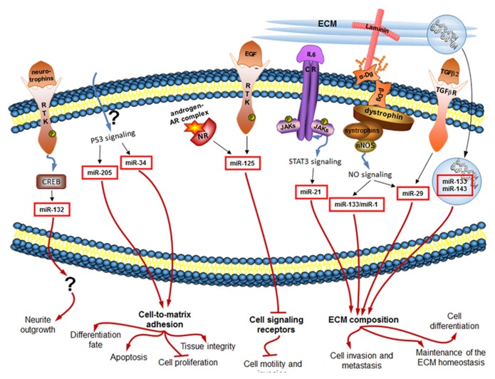
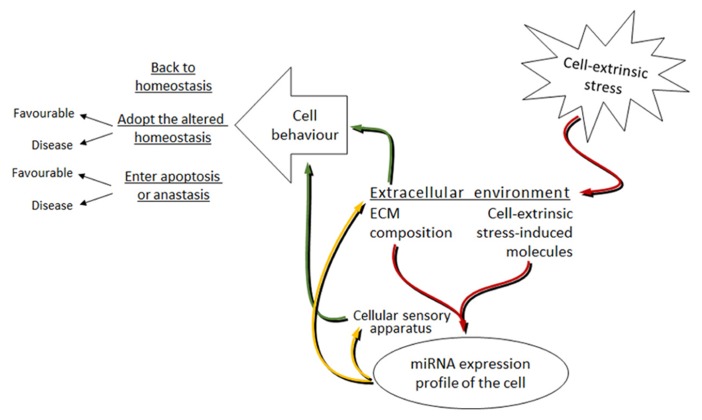
The extracellular environment is a complex entity comprising of the extracellular matrix (ECM) and regulatory molecules. It is highly dynamic and under cell-extrinsic stress, transmits the stressed organism's state to each individual ECM-connected cell. microRNAs (miRNAs) are regulatory molecules involved in virtually all the processes in the cell, especially under stress. In this review, we analyse how miRNA expression is regulated downstream of various signal transduction pathways induced by changes in the extracellular environment. In particular, we focus on the muscular dystrophy-associated cell adhesion molecule dystroglycan capable of signal transduction. Then we show how exactly the same miRNAs feedback to regulate the extracellular environment. The ultimate goal of this bi-directional signal transduction process is to change cell behavior under cell-extrinsic stress in order to respond to it accordingly.

Keywords: ECM composition; bi-directional signal transduction; dystroglycan; extrinsic stress; miRNAs.


Figures

FIGURE 1
FIGURE 1
The cell-extrinsic stress signaling brings about the changes in the extracellular environment that are read by the cellular sensory apparatus. The extracellular environment is a complex but organized entity composed of structural and enzymatic ECM proteins [collagens, proteoglycans1, multiadhesive proteins like laminin and fibronectin (Fn), MMPs and TIMPs] and cell-extrinsic stress-induced signaling molecules including cytokines (considering growth factors as a type of cytokine), hormones, and extracellular miRNAs. The cellular sensory apparatus includes the ECM receptors and receptors for signaling molecules. Integrins and dystroglycan (Dg) are the ECM receptors. They do not only connect cell to its matrix but are also involved in the cell signaling. Multiple receptors for signaling molecules [including receptor tyrosine kinases (RTK), cytokine receptors (CR), TGF-β receptors, and nuclear receptors (NR)] transduce changes in the extracellular environment into the various signaling pathways inside the cell. Extracellular miRNAs can probably be taken up by the cell via yet unknown mechanism. NO, nitric oxide, Ago, argonaute.
FIGURE 2
FIGURE 2
miRNAs regulated downstream of signaling induced by changes in the extracellular environment (black arrows) influence in turn the ECM composition and the cellular sensory apparatus, and ultimately the cell behavior (red arrows). Only mammalian miRNAs and pathways are shown.
FIGURE 3
FIGURE 3
Proposed model of the miRNAs involvement in the bi-directional signal transduction between the cell and extracellular environment under cell-extrinsic stress. Red arrows: cell-extrinsic stress changes the extracellular environment of cells in the organism. The ECM composition and the cell-extrinsic stress-induced molecules, as parts of the altered extracellular environment, signal via the cellular sensory apparatus to change the miRNA expression profile of cells. Yellow arrows: altered miRNA levels target the cellular sensory apparatus and the extracellular environment. Green arrows: this leads to the changes in cell behavior. The mode of cell behavior is different dependent on the strength and length of the applied cell-extrinsic stress.

Similar articles

Cited by

References

    1. Alessi A., Bragg A. D., Percival J. M., Yoo J., Albrecht D. E., Froehner S. C., et al. (2006).gamma-Syntrophin scaffolding is spatially and functionally distinct from that of the alpha/beta syntrophins. Exp. Cell Res. 312 3084–3095 10.1016/j.yexcr.2006.06.019 - DOI - PubMed
    1. Ariel A., Hershkoviz R., Cahalon L., Williams D. E., Akiyama S. K., Yamada K. M., et al. (1997). Induction of T cell adhesion to extracellular matrix or endothelial cell ligands by soluble or matrix-bound interleukin-7. Eur. J. Immunol. 27 2562–2570 10.1002/eji.1830271015 - DOI - PubMed
    1. Barresi R., Campbell K. P. (2006). Dystroglycan: from biosynthesis to pathogenesis of human disease. J. Cell Sci. 119 199–207 10.1242/jcs.02814 - DOI - PubMed
    1. Bhat H. F., Adams M. E., Khanday F. A. (2013). Syntrophin proteins as Santa Claus: role(s) in cell signal transduction. Cell. Mol. Life Sci. 70 2533–2554 10.1007/s00018-012-1233-9 - DOI - PMC - PubMed
    1. Bissell M. J., Hall H. G., Parry G. (1982). How does the extracellular matrix direct gene expression? J. Theor. Biol. 99 31–68 10.1016/0022-5193(82)90388-5 - DOI - PubMed

LinkOut - more resources