. 2014 Jan 10;9(1):e84777.
doi: 10.1371/journal.pone.0084777. eCollection 2014.

MR-less surface-based amyloid assessment based on 11C PiB PET

Collaborators, Affiliations

MR-less surface-based amyloid assessment based on 11C PiB PET

Luping Zhou et al. PLoS One. .

Abstract

Background: β-amyloid (Aβ) plaques in brain's grey matter (GM) are one of the pathological hallmarks of Alzheimer's disease (AD), and can be imaged in vivo using Positron Emission Tomography (PET) with (11)C or (18)F radiotracers. Estimating Aβ burden in cortical GM has been shown to improve diagnosis and monitoring of AD. However, lacking structural information in PET images requires such assessments to be performed with anatomical MRI scans, which may not be available at different clinical settings or being contraindicated for particular reasons. This study aimed to develop an MR-less Aβ imaging quantification method that requires only PET images for reliable Aβ burden estimations.

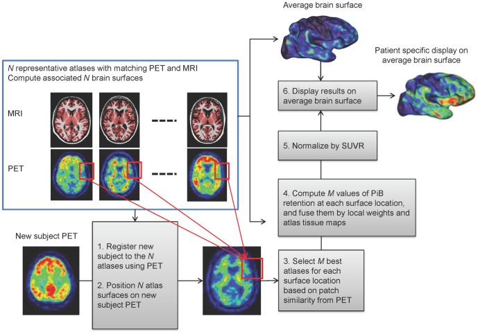
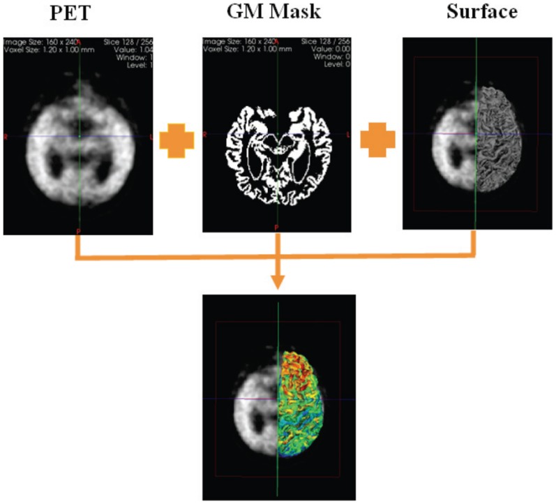
Materials and methods: The proposed method has been developed using a multi-atlas based approach on (11)C-PiB scans from 143 subjects (75 PiB+ and 68 PiB- subjects) in AIBL study. A subset of 20 subjects (PET and MRI) were used as atlases: 1) MRI images were co-registered with tissue segmentation; 2) 3D surface at the GM-WM interfacing was extracted and registered to a canonical space; 3) Mean PiB retention within GM was estimated and mapped to the surface. For other participants, each atlas PET image (and surface) was registered to the subject's PET image for PiB estimation within GM. The results are combined by subject-specific atlas selection and Bayesian fusion to generate estimated surface values.

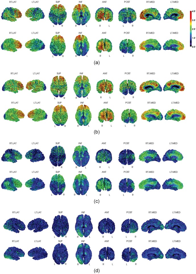
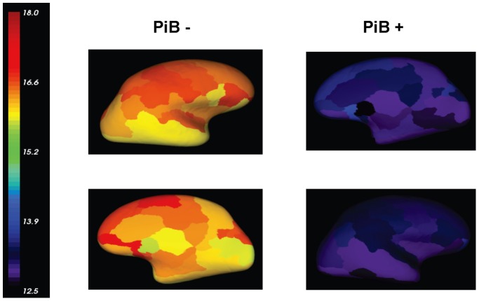
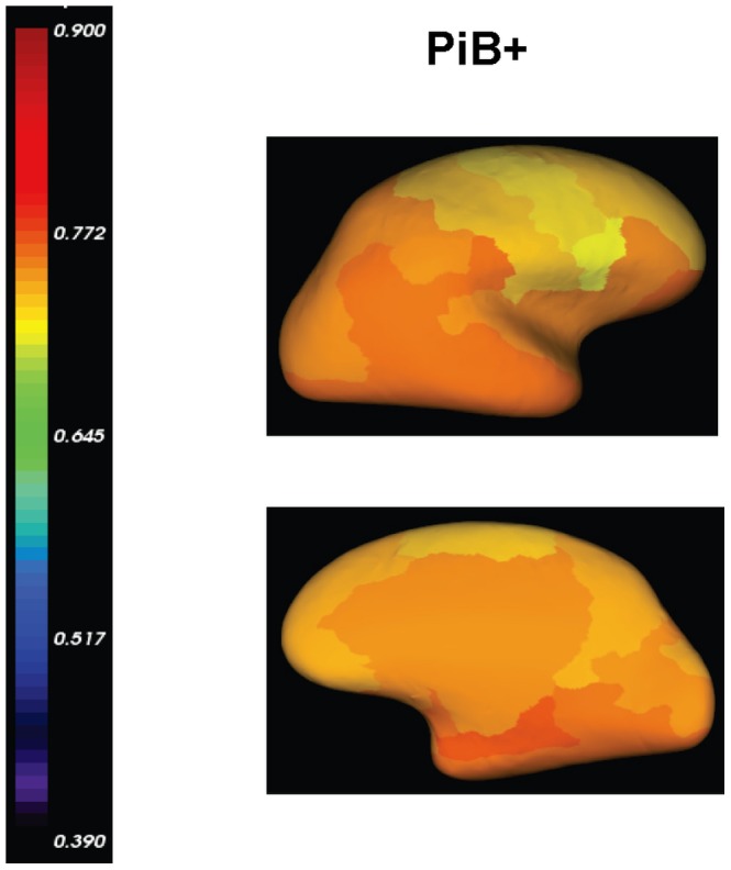
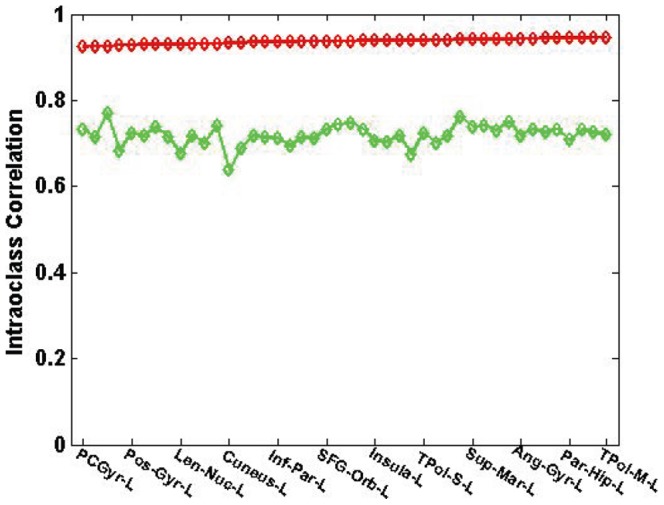
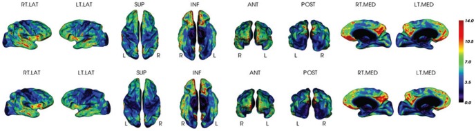
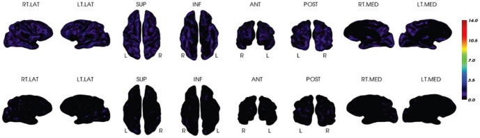
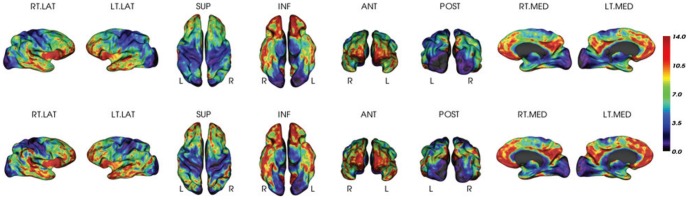
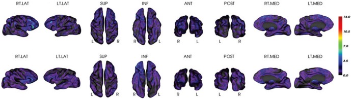
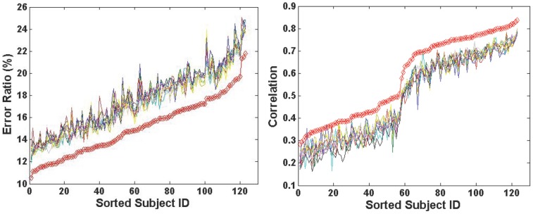
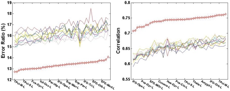
Results: All PiB+ subjects (N = 75) were highly correlated between the MR-dependent and the PET-only methods with Intraclass Correlation (ICC) of 0.94, and an average relative difference error of 13% (or 0.23 SUVR) per surface vertex. All PiB- subjects (N = 68) revealed visually akin patterns with a relative difference error of 16% (or 0.19 SUVR) per surface vertex.

Conclusion: The demonstrated accuracy suggests that the proposed method could be an effective clinical inspection tool for Aβ imaging scans when MRI images are unavailable.


Conflict of interest statement

Competing Interests: Luping Zhou, Olivier Salvado, Vincent Dore, Pierrick Bourgeat, Victor L. Villemagne, Christopher C. Rowe, Jurgen Fripp might be listed as co-inventor in a patent that has been lodged describing a technology using techniques described in this manuscript. The authors' patent application name is: Method and apparatus for the assessment of medical images. The patent application number is: PCT/AU2012/001536. More information could be found from this link: http://www.google.com/patents/WO2013086580A1?cl=en CogState Ltd. (http://cogstate.com/), Hollywood Private Hospital (http://www.hollywood.ramsayhealth.com.au/) and Sir Charles Gardner Hospital (http://www.scgh.health.wa.gov.au/) have contributed to the financial support of AIBL study through the partnership with the Science and Industry Endowment Fund (SIEF http://www.sief.org.au/). Pfizer International (http://www.pfizer.com.au/default.aspx) has contributed financial support to AIBL to assist with analysis of blood samples and to further the AIBL research program. There are no further patents, products in development or marketed products to declare. This does not alter the authors' adherence to all the PLOS ONE policies on sharing data and materials, as detailed online in the guide for authors.

Figures

Figure 1
Figure 1. Overview of the proposed method.
Figure 2
Figure 2. Illustration of the MRI-dependent method.
The PiB retention is measured in the PET image within its grey matter mask obtained from MRI tissue segmentation, and averaged along the normal direction of the GM-WM interface (overlaid on the PET image) extracted from the subject's MRI. The mean PiB value for each surface vertex is mapped onto the surface for visualization.
Figure 3
Figure 3. Visual Inspection for PiB measurements.
Surface-based PiB measurements from the MRI-dependent method (the top row) and the proposed method (the bottom row) for four examples: (a) AD, (b) PiB+ NC, (c) PiB+ NC, and (d) PiB- NC.
Figure 4
Figure 4. Vertex-based mean estimation errors (ratio) in each AAL ROI.
The errors are visualized on an inflated template brain surface for both PiB+ and PiB- groups. There are higher estimation error ratios for PiB- group than for PiB+ group, due to the minimal amount of retention and the reduced dynamic range of PiB- group. The mean absolute estimation error (vertex-based) is 0.19±0.03 for PiB- group and 0.23±0.04 for PiB+ group as reported in Table 3 and Table 4.
Figure 5
Figure 5. Mean correlations between PET-only and MRI-dependent methods over AAL ROIs.
The correlations are visualized on inflated template brain surface for both PiB+ and PiB- groups.
Figure 6
Figure 6. Intra-class correlation over AAL ROIs between PET-only and the MRI-dependent methods.
Red line is for the PiB+ group and green line is for the PiB- group. To improve clarity, not all ROI names are shown in the graph.
Figure 7
Figure 7. Mean Z-score for PiB+ AD group.
The Z-scores are estimated by the MRI-dependent (top row) and the PET-only (bottom row) methods, respectively.
Figure 8
Figure 8. Mean Z-score for PiB- NC group.
The Z-scores are estimated by the MRI-dependent (top row) and the PET-only (bottom row) methods, respectively.
Figure 9
Figure 9. Z-score for an individual PiB+ subject.
It is estimated by the MRI-dependent (top row) and the PET-only (bottom row) methods, respectively.
Figure 10
Figure 10. Z-score for an individual PiB- subject.
It is estimated by the MRI-dependent (top row) and the PET-only (bottom row) methods, respectively.
Figure 11
Figure 11. Comparison between the multiple-atlas and ten single-atlas approaches subject by subject (left: error ratio; right: correlation).
The red line corresponds to the proposed multiple-atlas approach, while the rest ten lines correspond to the ten single-atlas approaches in comparison, respectively. To improve clarity, the subjects' IDs are sorted according to the increase of error ratios and correlation, respectively.
Figure 12
Figure 12. Comparison of the multiple-atlas and ten single-atlas approaches over AAL ROIs within PiB+ group (left: error ratio; right: correlation).
The red line corresponds to the proposed multiple-atlas approach, while the rest ten lines correspond to the ten single-atlas approaches in comparison, respectively. To improve clarity, not all ROI names are shown in the graph.
Figure 13
Figure 13. Comparison of the multiple-atlas and ten single-atlas approaches over AAL ROIs within PiB- group (left: error ratio; right: correlation).
The red line corresponds to the proposed multiple-atlas approach, while the rest ten lines correspond to the ten single-atlas approaches in comparison, respectively. To improve clarity, not all ROI names are shown in the graph.

Similar articles

Cited by

References

    1. Aalto S, Scheinin MN, Kemppainen MN, Någren K, Kailajärvi M, et al. (2009) Reproducibility of automated simplified voxel-based analysis of PET amyloid ligand [11C]PIB uptake using 30-min scanning data. Eur J Nucl Med Mol Imaging 36: 1651–1660. - PubMed
    1. Acosta O, Fripp J, Doré V, Bourgeat P, Favreau JM, et al. (2012) Cortical surface mapping using topology correction, partial flattening and 3D shape context-based non-rigid registration for use in quantifying atrophy in Alzheimer's disease. Journal of Neuroscience Methods 205: 96–109. - PubMed
    1. Artaechevarria X, Munoz-Barrutia A, Ortiz-de-Solorzano C (2009) Combination strategies in multi-atlas image segmentation: application to brain MR data. IEEE Trans Med Imaging 28 (8): 1266–1277. - PubMed
    1. Bourgeat P, Raniga P, Dore V, Zhou L, Macaulay SL, et al... (2012) Manifold Driven MR-less PiB SUVR Normalisation. In MICCAI 2012 Workshop on Novel Imaging Biomarkers for Alzheimer's Disease and Related Disorders (NIBAD'12)
    1. Braak H, Braak E (1991) Neuropathological stageing of Alzheimer-related changes. Acta Neuropathol. (Berl) 82 (4): 239–259. - PubMed

Publication types