Role of PDGFs/PDGFRs signaling pathway in myocardial fibrosis of DOCA/salt hypertensive rats
- PMID: 24427322
- PMCID: PMC3885456
Role of PDGFs/PDGFRs signaling pathway in myocardial fibrosis of DOCA/salt hypertensive rats
Abstract
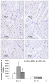
This study aimed to investigate the role of PDGF/PDGFR signaling pathway in myocardial fibrosis of desoxycorticosterone (DOCA) induced salt-sensitive hypertensive rats and explore the influence of PDGF/PDGFR signaling pathway on fibroblasts and myofibroblasts in the heart. 60 male SD rats underwent right nephrectomy and bred with 1% sodium chloride and 0.1% potassium chloride for 4 weeks, and then randomly divided into 3 groups (CON group, DOCA group and DOCA+IMA group). Results showed that: 1) 14 and 28 days after intervention, the SBP in DOCA and DOCA+IMA group was significantly higher than that in CON group. At days 28, the severity of myocardial fibrosis and PVCA/VA ratio in DOCA group were significantly increased when compared with CON group. The severity of myocardial fibrosis and PVCA/VA ratio in DOCA+IMA group were markedly lower than those in DOCA group although they were higher than those in CON group. 2) At days 14, the mRNA expressions of PDGFRα and PDGFRβ in DOCA group were significantly higher than CON and DOCA+IMA group. At days 28, the mRNA expressions of PDGFRβ, FSP-1, α-SMA, procollagen I and procollagen III in DOCA group were significantly higher than those in CON group. In addition, in a specific group, the PDGFRβ mRNA expression was higher than the PDGFRα mRNA expression. In DOCA+IMA group, the mRNA expressions of PDGFRβ, FSP-1, α-SMA, procollagen I and procollagen III were markedly reduced when compared with DOCA group. 3) At 14 days, the protein expressions of PDGFRα and PDGFRβ in DOCA group were significantly higher than those in CON group. The PDGFRα protein expression in DOCA+IMA group was markedly lower than that in DOCA group. At days 28, the protein expressions of PDGFRα and PDGFRβ in DOCA group were significantly increased when compared with CON group. The protein expressions of PDGFRα and PDGFRβ in DOCA+IMA group were significantly lower than those in DOCA group. At day 28, the cardiac interstitium mainly contained vimentin positive fibroblasts, and α-SMA positive cells were less identified in CON group. In DOCA group, α-SMA positive fibroblasts (spindle-shaped) increased significantly, but the myofibroblasts reduced significantly in DOCA+IMA group when compared with DOCA group. 4) PDGFRα protein expression was observed in fibroblasts and myofibroblasts, but not in VSMCs. PDGFRβ protein expression was noted in not only fibroblasts and myofibroblasts but also VSMCs. Thus, During myocardial fibrosis of DOCA induced salt-sensitive hypertensive rats, PDGFRα acts at early stage, but PDGFRβ functions in the whole process. PDGFRα and PDGFRβ expressions increase in fibroblasts and myofibroblasts, suggesting that PDGF/PDGFR signaling pathway is involved in the myocardial fibrosis via stimulating fibroblasts to proliferate and transform into myofibroblasts.
Keywords: Platelet-derived growth factor; desoxycorticosterone; fibroblasts; imatinib; myocardial fibrosis; myofibroblasts; platelet-derived growth factor receptor.
Figures


References
-
- Zeisberg EM, Tarnavski O, Zeisberg M, Dorfman AL, McMullen JR, Gustafsson E, Chandraker A, Yuan X, Pu WT, Roberts AB, Neilson EG, Sayegh MH, Izumo S, Kalluri R. Endothelial-to-mesenchymal transition contributes to cardiac fibrosis. Nat Med. 2007;13:952–961. - PubMed
-
- Ma LK, Li Q, He LF, Hua JS, Zhou JL, Yu H, Feng KF, Chen HW, Hu H, Wang L. Imatinib attenuates myocardial fibrosis in association with inhibition of the PDGFRalpha activity. Arq Bras Cardiol. 2012;99:1082–1091. - PubMed
-
- Yu M, Ishibashi-Ueda H, Ohta-Ogo K, Gabbiani G, Yamagishi M, Hayashi K, Hirota S, Bochaton-Piallat ML, Hao H. Transient expression of cellular retinol-binding protein-1 during cardiac repair after myocardial infarction. Pathol Int. 2012;62:246–253. - PubMed
-
- Zymek P, Bujak M, Chatila K, Cieslak A, Thakker G, Entman ML, Frangogiannis NG. The role of platelet-derived growth factor signaling in healing myocardial infarcts. J Am Coll Cardiol. 2006;48:2315–2323. - PubMed
Publication types
MeSH terms
Substances
LinkOut - more resources
Full Text Sources
Medical
Research Materials
Miscellaneous
