

Correlation of histological and macroscopic findings in peritoneal endometriosis
- PMID: 24427335
- PMCID: PMC3885469
Correlation of histological and macroscopic findings in peritoneal endometriosis
Abstract
Context: In the last two decades, a color based concept of disease activity in peritoneal endometriosis has been in use in the clinical context, with red lesions being considered active and black or white lesions being interpreted as less active or dormant.
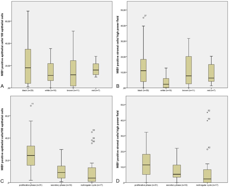
Objective: Our aim was to analyze 4 main color categories of peritoneal endometriosis (black, white, red and brown) in one single patient group using histomorphological and immunohistochemical methods.
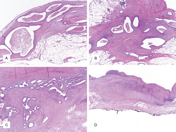
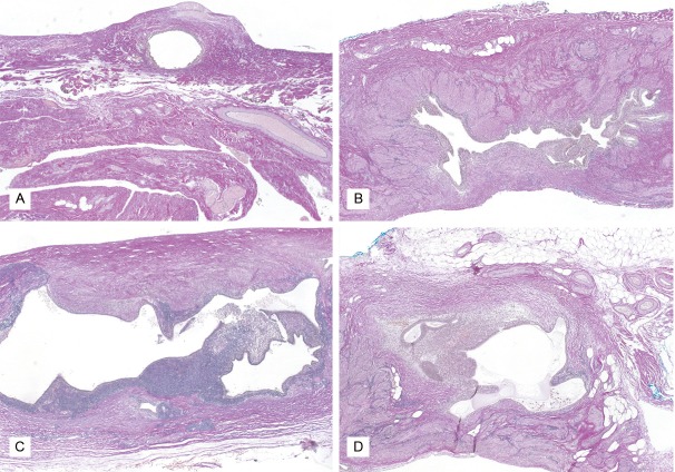
Design: 65 endometriosis lesions (30 black, 17 white, 11 brown, 7 red) were resected from 47 premenopausal, nulliparous women which had not received exogenous hormones for at least six months prior to the operation. Specimen workup, histomorphological analysis and immunohistochemical analysis were performed in a standardized manner.
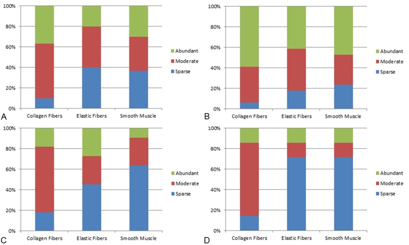
Results: The color categories showed a broad overlap in proliferative activity and hormone receptor expression. Differences were found in lesion morphology. Adjacent stromal reaction in particular showed a marked increase from red through brown and black to white lesions. Differences were also seen in gland pattern and gland content.
Conclusions: Lesion colors in peritoneal endometriosis seem to be determined by gland content and a varying adjacent stromal reaction and more likely reflect an aging process than different levels of disease activity.
Keywords: Endometriosis; age; colors; hormone receptors; proliferation.
Figures
References
-
- Renner SP, Lermann J, Hackl J, Burghaus S, Oppelt P, Binder H. Chronische Erkrankung. Endometriose. Geburtsh Frauenheilk. 2012;72:914–919.
-
- Droegemueller W. Comprehensive Gynecology. Philadelphia: Mosby; 2001.
-
- Rokitansky C. Ueber Uterusdruesen-Neubildung in Uterus und Ovarialsarcomen. Z Ges Aerzte Wien. 1860;16:577–581.
-
- Sampson JA. Peritoneal endometriosis due to menstrual dissemination of endometrial tissue into the peritoneal cavity. Am J Obstet Gynecol. 1927;14:422.
-
- Donnez J, Squifflet J, Casanas-Roux F, Pirard C, Jadoul P, Van Langendonckt A. Typical and subtle atypical presentations of endometriosis. Obstet Gynecol Clin North Am. 2003;30:83–93. viii. - PubMed
Publication types
MeSH terms
LinkOut - more resources
Full Text Sources
Other Literature Sources
Medical