

Fungus balls of the bilateral paranasal sinuses
- PMID: 24427669
- PMCID: PMC3738784
- DOI: 10.1007/s12070-011-0465-6
Fungus balls of the bilateral paranasal sinuses
Abstract
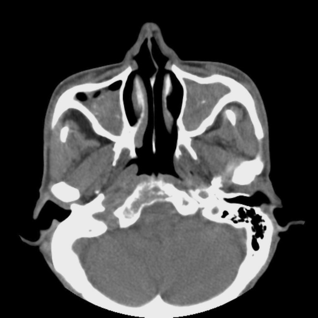
We describe the clinical and radiographic characteristics of fungus balls in the bilateral paranasal sinuses. The medical records of 8 of 245 patients with fungus balls of the bilateral paranasal sinuses between 2000 and 2010 were retrospectively reviewed. The incidence of bilateral paranasal sinus fungus balls was 3.3%. Fungus balls were located in the maxillary sinuses bilaterally in 4 cases (50%), followed by the maxillary sinus and contralateral sphenoid sinus in 3 cases (37.5%), and the sphenoid sinuses bilaterally in 1 case (12.5%). There were no predisposing anatomic variations for the occurrence of bilateral paranasal fungus balls. Although the presenting symptoms and signs were non-specific, CT findings were helpful in the diagnosis of bilateral fungus balls. Endonasal removal by an endoscopic approach was performed in all patients. No peri-operative complications or recurrences were noted. Fungus balls in the bilateral paranasal sinuses are most frequently found in the maxillary sinuses bilaterally. Because symptoms of bilateral paranasal fungus balls and findings on nasal endoscopic examination are frequently non-specific, a high index of suspicion is needed and imaging studies, such as CT, are essential to establish the correct pre-operative diagnosis.
Keywords: Bilateral; Endoscopic surgical procedure; Fungus disease; Paranasal sinuses; Sinusitis.
Figures
Similar articles
-
Paranasal sinus fungus balls.Head Neck. 1997 Sep;19(6):481-6. doi: 10.1002/(sici)1097-0347(199709)19:6<481::aid-hed4>3.0.co;2-v. Head Neck. 1997. PMID: 9278755
-
Triple discrete fungus balls of the paranasal sinuses.Otolaryngol Head Neck Surg. 2004 Dec;131(6):1014-5. doi: 10.1016/j.otohns.2004.02.013. Otolaryngol Head Neck Surg. 2004. PMID: 15577809
-
There is no structural relationship between nasal septal deviation, concha bullosa, and paranasal sinus fungus balls.ScientificWorldJournal. 2012;2012:181246. doi: 10.1100/2012/181246. Epub 2012 Dec 26. ScientificWorldJournal. 2012. PMID: 23326212 Free PMC article.
-
Fungus balls of the paranasal sinuses: a review.Eur Arch Otorhinolaryngol. 2007 May;264(5):461-70. doi: 10.1007/s00405-007-0281-5. Epub 2007 Mar 15. Eur Arch Otorhinolaryngol. 2007. PMID: 17361410 Review.
-
Fungal diseases of the sinuses.Otolaryngol Head Neck Surg. 1990 Dec;103(6):1012-5. doi: 10.1177/019459989010300621. Otolaryngol Head Neck Surg. 1990. PMID: 2126115 Review.
Cited by
-
Comparison of the clinical characteristics of bilateral and unilateral fungal balls in Korea.Eur Arch Otorhinolaryngol. 2019 Jul;276(7):1975-1980. doi: 10.1007/s00405-019-05408-6. Epub 2019 Mar 30. Eur Arch Otorhinolaryngol. 2019. PMID: 30929057
-
Clinical features of chronic fungal rhinosinusitis in Korean geriatric and non-geriatric patients.Eur Arch Otorhinolaryngol. 2023 Nov;280(11):4969-4977. doi: 10.1007/s00405-023-08089-4. Epub 2023 Jun 30. Eur Arch Otorhinolaryngol. 2023. PMID: 37389593
-
An unusual presentation of sphenoid Candida fungal ball: a case report.J Surg Case Rep. 2019 Aug 12;2019(8):rjz240. doi: 10.1093/jscr/rjz240. eCollection 2019 Aug. J Surg Case Rep. 2019. PMID: 31423297 Free PMC article.
-
The Endonasal Endoscopic Approach to Different Sinonasal Fungal Balls.Int J Otolaryngol. 2022 Mar 22;2022:6721896. doi: 10.1155/2022/6721896. eCollection 2022. Int J Otolaryngol. 2022. PMID: 35360416 Free PMC article.
-
Isolated sphenoid sinus fungus ball: a retrospective study conducted at a tertiary care referral center in Korea.Eur Arch Otorhinolaryngol. 2017 Jun;274(6):2453-2459. doi: 10.1007/s00405-017-4468-0. Epub 2017 Mar 1. Eur Arch Otorhinolaryngol. 2017. PMID: 28251318
References
-
- Dufour X, Kauffmann-Lacroix C, Ferrie JC, Goujon JM, Rodier MH, Karkas A, et al. Paranasal sinus fungus ball and surgery: a review of 175 cases. Rhinology. 2005;43:34–39. - PubMed
LinkOut - more resources
Full Text Sources