

Efficacy assessment of a combined anticholinergic and oxime treatment against topical sarin-induced miosis and visual impairment in rats
- PMID: 24428128
- PMCID: PMC3997276
- DOI: 10.1111/bph.12586
Efficacy assessment of a combined anticholinergic and oxime treatment against topical sarin-induced miosis and visual impairment in rats
Abstract
Background and purpose: Eye exposure to the organophosphorus (OP) irreversible cholinesterase inhibitor sarin results in long-term miosis and impaired visual function. We have previously shown that tropicamide is better at ameliorating this insult than topical atropine or cyclopentolate. However, to minimize side effects associated with repeated tropicamide applications and high treatment doses, we evaluated the effects of oximes (ChE re-activators) alone and combined with tropicamide at ameliorating OP-induced ocular impairments.
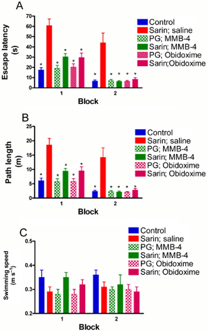
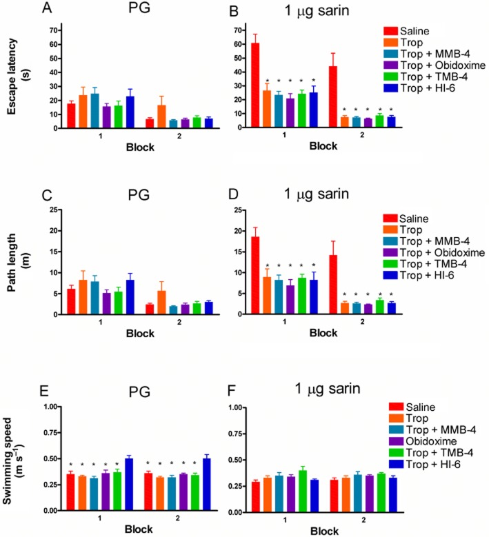
Experimental approach: Rats were topically exposed to sarin, followed by topical treatment with various oximes alone or in combination with tropicamide. Pupil width and light reflex were measured by an infrared-based digital photograph system, while visual performance was assessed by employing the cueing version of the Morris water maze (MWM).
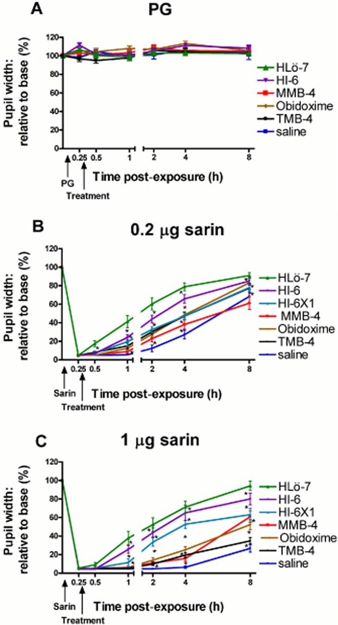
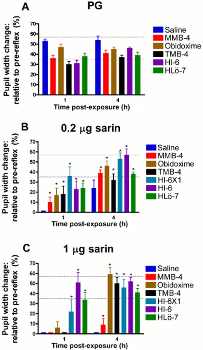
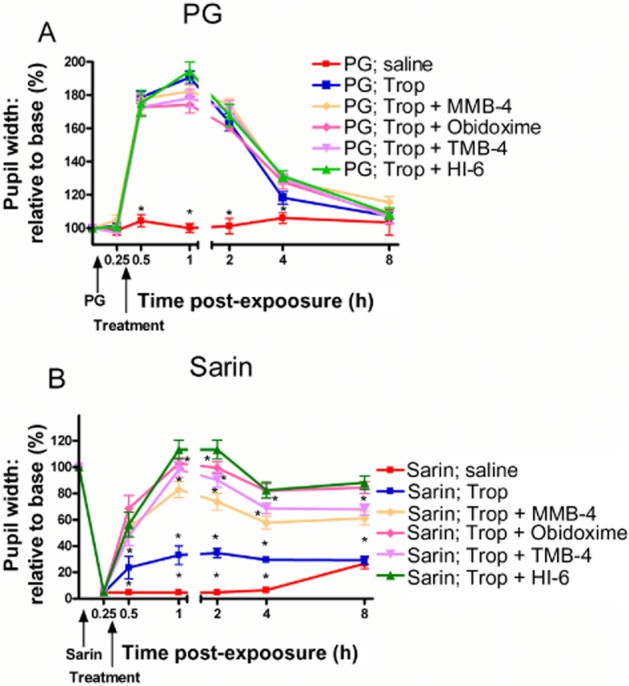
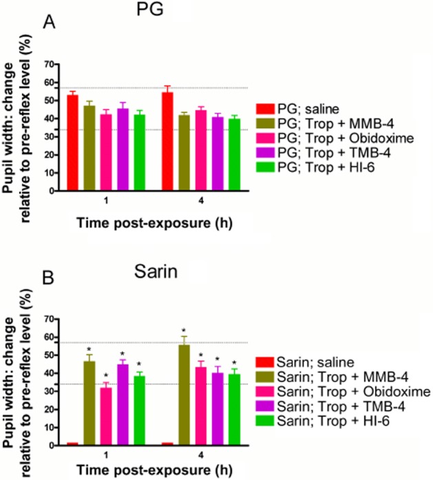
Key results: Oxime treatment following sarin ocular exposure induced a slow persistent pupil widening with efficacy in the order of HLö-7 > HI-6 > obidoxime = TMB-4 = MMB-4. In the light reflex test, the ability of the iris to contract following oxime treatment was mostly impaired at 1 h and was back to normal at 4 h following sarin exposure. All oxime treatments ameliorated the sarin-induced visual impairment as tested in the visual task (MWM). The combined topical treatment of tropicamide with an oxime induced a rapid improvement in pupil widening, light reflex and visual performance, and enabled a reduction in tropicamide dose.
Conclusions and implications: The use of tropicamide combined with an oxime should be considered as the topical treatment of choice against the toxic effects of ocular OP exposure.
Keywords: HI-6; HLö-7; Long-Evans rats; MMB-4; TMB-4; anticholinergic treatments; cued MWM; miosis; obidoxime; organophosphates; oxim; pupillary light reflex; sarin; tropicamide; visual impairment.
© 2014 The British Pharmacological Society.
Figures
Similar articles
-
Synergism Between Anticholinergic and Oxime Treatments Against Sarin-Induced Ocular Insult in Rats.Toxicol Sci. 2015 Aug;146(2):301-10. doi: 10.1093/toxsci/kfv092. Epub 2015 May 7. Toxicol Sci. 2015. PMID: 25956921
-
Efficacy assessment of various anticholinergic agents against topical sarin-induced miosis and visual impairment in rats.Toxicol Sci. 2012 Apr;126(2):515-24. doi: 10.1093/toxsci/kfs009. Epub 2012 Jan 12. Toxicol Sci. 2012. PMID: 22247005
-
Optimization of the Ocular Treatment Following Organophosphate Nerve Agent Insult.Toxicol Sci. 2017 Sep 1;159(1):50-63. doi: 10.1093/toxsci/kfx119. Toxicol Sci. 2017. PMID: 28903494
-
Broad Spectrum Treatment for Ocular Insult Induced by Organophosphate Chemical Warfare Agents.Toxicol Sci. 2020 Sep 1;177(1):1-10. doi: 10.1093/toxsci/kfaa095. Toxicol Sci. 2020. PMID: 32579218 Review.
-
Unequal efficacy of pyridinium oximes in acute organophosphate poisoning.Clin Med Res. 2007 Mar;5(1):71-82. doi: 10.3121/cmr.2007.701. Clin Med Res. 2007. PMID: 17456837 Free PMC article. Review.
Cited by
-
Eyeing up the Future of the Pupillary Light Reflex in Neurodiagnostics.Diagnostics (Basel). 2018 Mar 13;8(1):19. doi: 10.3390/diagnostics8010019. Diagnostics (Basel). 2018. PMID: 29534018 Free PMC article. Review.
References
-
- Bartlett JD, Jaanus SD, Fiscella RG, Holdeman NR, Prokopich CL. Clinical Ocular Pharmacology. 5. St. Louis: Elsevier; 2008a. Chapter Nine.
-
- Bartlett JD, Jaanus SD, Fiscella RG, Holdeman NR, Prokopich CL. Clinical Ocular Pharmacology. 5. St. Louis: Elsevier; 2008b. Chapter Two.
-
- Brandeis R, Brandys Y, Yehuda S. The use of the Morris Water Maze in the study of memory and learning. Int J Neurosci. 1989;48:29–69. - PubMed
-
- Cannard K. The acute treatment of nerve agent exposure. J Neurol Sci. 2006;249:86–94. - PubMed
MeSH terms
Substances
LinkOut - more resources
Full Text Sources
Other Literature Sources
Medical
Miscellaneous
