

Disrupting the Indian hedgehog signaling pathway in vivo attenuates surgically induced osteoarthritis progression in Col2a1-CreERT2; Ihhfl/fl mice
- PMID: 24428864
- PMCID: PMC3978435
- DOI: 10.1186/ar4437
Disrupting the Indian hedgehog signaling pathway in vivo attenuates surgically induced osteoarthritis progression in Col2a1-CreERT2; Ihhfl/fl mice
Abstract
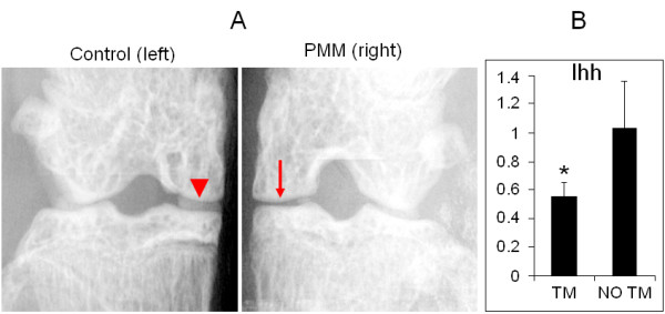
Introduction: Previous observations implicate Indian hedgehog (Ihh) signaling in osteoarthritis (OA) development because it regulates chondrocyte hypertrophy and matrix metallopeptidase 13 (MMP-13) expression. However, there is no direct genetic evidence for the role of Ihh in OA, because mice with cartilage or other tissue-specific deletion of the Ihh gene die shortly after birth. We evaluated the role of Ihh in vivo via a Cre-loxP-mediated approach to circumvent the early death caused by Ihh deficiency.
Methods: To evaluate the role of Ihh in OA development, Ihh was specifically deleted in murine cartilage using an Ihh conditional deletion construct (Col2a1-CreER(T2); Ihh(fl/fl)). The extent of cartilage degradation and OA progression after Ihh deletion was assessed by histological analysis, immunohistochemistry, real-time PCR and in vivo fluorescence molecular tomography (FMT) 2 months after OA was induced by partial medial meniscectomy. The effect of Ihh signaling on cartilage was compared between Ihh-deleted mice and their control littermates.
Results: Only mild OA changes were observed in Ihh-deleted mice, while control mice displayed significantly more cartilage damage. Typical OA markers such as type X collagen and MMP-13 were decreased in Ihh-deleted mice. In vivo FMT demonstrated decreased cathepsins and MMP activity in knee joints of animals with deletion of Ihh.
Conclusions: These findings support the protective role of Ihh deletion in surgically induced OA. Thus, our findings suggest the potential to develop new therapeutic strategies that can prevent and treat OA by inhibiting Ihh signaling in chondrocytes.
Figures


Similar articles
-
Indian Hedgehog, a critical modulator in osteoarthritis, could be a potential therapeutic target for attenuating cartilage degeneration disease.Connect Tissue Res. 2014 Aug;55(4):257-61. doi: 10.3109/03008207.2014.925885. Epub 2014 Jun 13. Connect Tissue Res. 2014. PMID: 24844414 Review.
-
Activation of Indian hedgehog promotes chondrocyte hypertrophy and upregulation of MMP-13 in human osteoarthritic cartilage.Osteoarthritis Cartilage. 2012 Jul;20(7):755-63. doi: 10.1016/j.joca.2012.03.010. Epub 2012 Mar 30. Osteoarthritis Cartilage. 2012. PMID: 22469853 Free PMC article.
-
Ipriflavone attenuates the degeneration of cartilage by blocking the Indian hedgehog pathway.Arthritis Res Ther. 2019 May 2;21(1):109. doi: 10.1186/s13075-019-1895-x. Arthritis Res Ther. 2019. PMID: 31046827 Free PMC article.
-
MicroRNA‑1 regulates the development of osteoarthritis in a Col2a1‑Cre‑ERT2/GFPfl/fl‑RFP‑miR‑1 mouse model of osteoarthritis through the downregulation of Indian hedgehog expression.Int J Mol Med. 2020 Jul;46(1):360-370. doi: 10.3892/ijmm.2020.4601. Epub 2020 May 12. Int J Mol Med. 2020. PMID: 32626917 Free PMC article.
-
Functional role of hedgehog pathway in osteoarthritis.Cell Biochem Funct. 2020 Mar;38(2):122-129. doi: 10.1002/cbf.3448. Epub 2019 Dec 12. Cell Biochem Funct. 2020. PMID: 31833076 Review.
Cited by
-
Signalling interaction between β-catenin and other signalling molecules during osteoarthritis development.Cell Prolif. 2024 Jun;57(6):e13600. doi: 10.1111/cpr.13600. Epub 2024 Jan 10. Cell Prolif. 2024. PMID: 38199244 Free PMC article. Review.
-
siRNA-based nanotherapeutics as emerging modalities for immune-mediated diseases: A preliminary review.Cell Biol Int. 2022 Sep;46(9):1320-1344. doi: 10.1002/cbin.11841. Epub 2022 Jul 13. Cell Biol Int. 2022. PMID: 35830711 Free PMC article. Review.
-
Cat's whiskers (Orthosiphon stamineus) tea modulates arthritis pathogenesis via the angiogenesis and inflammatory cascade.BMC Complement Altern Med. 2016 Nov 24;16(1):480. doi: 10.1186/s12906-016-1467-4. BMC Complement Altern Med. 2016. PMID: 27881135 Free PMC article.
-
Attenuation of cartilage pathogenesis in post-traumatic osteoarthritis (PTOA) in mice by blocking the stromal derived factor 1 receptor (CXCR4) with the specific inhibitor, AMD3100.J Orthop Res. 2015 Jul;33(7):1071-8. doi: 10.1002/jor.22862. Epub 2015 Apr 24. J Orthop Res. 2015. PMID: 25732515 Free PMC article.
-
Hedgehog Signalling Contributes to Trauma-Induced Tendon Heterotopic Ossification and Regulates Osteogenesis through Antioxidant Pathway in Tendon-Derived Stem Cells.Antioxidants (Basel). 2022 Nov 16;11(11):2265. doi: 10.3390/antiox11112265. Antioxidants (Basel). 2022. PMID: 36421451 Free PMC article.
References
Publication types
MeSH terms
Substances
Grants and funding
LinkOut - more resources
Full Text Sources
Other Literature Sources
Medical
Molecular Biology Databases
