. 2014 Jan 15:13:16.
doi: 10.1186/1476-511X-13-16.

LDL-cholesterol signaling induces breast cancer proliferation and invasion

Affiliations

LDL-cholesterol signaling induces breast cancer proliferation and invasion

Catarina Rodrigues dos Santos et al. Lipids Health Dis. .

Abstract

Lipids and cholesterol in particular, have long been associated with breast cancer (BC) onset and progression. However, the causative effects of elevated lipid levels and breast cancer remain largely undisclosed and were the subject of the present study.We took advantage of well-established in vitro and in vivo models of cholesterol enrichment to exploit the mechanism involved in LDL-cholesterol favouring BC growth and invasiveness. We analyzed its effects in models that mimic different BC subtypes and stages.Our data show that LDL-cholesterol (but not HDL-cholesterol) promotes BC cells proliferation, migration and loss of adhesion, hallmarks of the epithelial to mesenchymal transition. In vivo studies modeling cholesterol levels showed that breast tumors are consistently larger and more proliferative in hypercholesterolemic mice, which also have more frequently lung metastases. Microarray analysis revealed an over expression of intermediates of Akt and ERK pathways suggesting a survival response induced by LDL, confirmed by WB analyses. Gene expression analysis also evidenced an activation of ErbB2 signaling pathway and decreased expression of adhesion molecules (cadherin-related family member3, CD226, Claudin 7 and Ocludin) in the cells exposed to LDL.Together, the present work shows novel mechanistic evidence that high LDL-cholesterol levels promote BC progression. These data provide rationale for the clinical control of cholesterol levels in BC patients.


Figures

Figure 1
Figure 1
Proliferation, migration and loss of adhesion induced by LDL in in breast cancer cell lines. A, Number of cells (MDA MB 231, HTB 126, HTB 20) after 24 h and 48 h of LDL (100 μg/mg) exposure is consistently higher than control (non-treated) condition. B and C, Cells (MDA MB 231) were wounded and then cultured for 24 h, with LDL (100 μg/mL), HDL (100 μg/mL) or control conditions. Cell migration into the wound was examined by phase-contrast microscopy and migration distance is indicated as the percentage of the wound closure at 24 h. Representative photos are shown (original magnification 100x C). D and E, Number of cells (MDA MB 231) adherent and no adherent cells, at 4 h after being removed and reseeded on its primary conditions (control and LDL100 μg/mL) shows that LDL treated cells lose matrix adhesion compared to control. Representative photos are shown (original magnification 200x E). P value and the number of the experiments are represented in the figure. Columns mean; bars ± SEM.
Figure 2
Figure 2
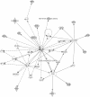
Activated cellular networks, at 48 h, in LDL treated breast cancer cell line. Gene expression analysis of breast cancer cells MDA MB231 exposed to LDL (100 μg/mL), for 48 h shows up regulation of molecules involved in activation of ERK, akt and ErbB2 pathways. Grey nodes are genes overexpressed, white nodes are predicted genes. Smooth lines represent direct interactions. Dashed lines represent indirect interactions.
Figure 3
Figure 3
LDL induces ERK and akt protein phosphorylation. A. Cells (MDA MB 231) exposed to LDL show higher expression of phosphorylated ERK and akt, without significant increase in respective total protein. Phosphorilation of JNK is also higher, without reaching statistical significance. Epidermal growth factor (EGF) stimulation was used as positive control and induced, as expected, increase ERK, akt and JNK phosphorylation. B. Representative photos of western blot membranes are shown. P value and the number of the experiments are represented in the figure. Columns mean; bars ± SEM.
Figure 4
Figure 4
Hypercholesterolemic diet induces a breast cancer phenotype characterized by large and more proliferative tumors. A and B. HD fed mice have raised levels of total cholesterol (TC), low density lipoprotein (LDL) and high density lipoprotein (HDL). There are no statistical significant differences in triglycerides level or animal weight, as exemplified to MDA MB 231/BALB SCID trial. (Results of the other experiments are in supplementary data.) Animal in the same diet but without tumor cells inoculation were used as control to lipid profile parameters. C and D. HD fed mice show large tumors when compared to ND fed mice. The mammary tumor large diameter was measured (as exemplified to 4 T1 /NOD experiment B) and the differences are shown as the fold change over the ND fed mice. E. HD fed mice are more likely to have lung metastasis (100%) than ND fed mice (33%). F and G. HD fed mice have more proliferative tumors as confirmed by Ki67 immuno staining. P value and the number of the experiments are represented in the figure. Columns mean; bars ± SEM. Scale bar 100 μm.
Figure 5
Figure 5
Phenotype of breast tumors exposed to high levels of cholesterol-Proposed Model. Breast tumors exposed to high levels of LDL-cholesterol are larger and show increased proliferation and migration while losing epithelial adhesion properties.

References

    1. Lingwood D, Simons K. Lipid rafts as a membrane-organizing principle. Science. 2010;327(5961):46–50. doi: 10.1126/science.1174621. - DOI - PubMed
    1. Menendez JA, Lupu R. Fatty acid synthase and the lipogenic phenotype in cancer pathogenesis. Nat Rev Cancer. 2007;7(10):763–777. doi: 10.1038/nrc2222. - DOI - PubMed
    1. Nomura DK, Long JZ, Niessen S, Hoover HS, Ng SW, Cravatt BF. Monoacylglycerol lipase regulates a fatty acid network that promotes cancer pathogenesis. Cell. 2010;140(1):49–61. doi: 10.1016/j.cell.2009.11.027. - DOI - PMC - PubMed
    1. Antalis CJ, Uchida A, Buhman KK, Siddiqui RA. Migration of MDA-MB-231 breast cancer cells depends on the availability of exogenous lipids and cholesterol esterification. Clin Exp Metastasis. 2011;28(8):733–741. doi: 10.1007/s10585-011-9405-9. - DOI - PubMed
    1. Swyer G. The cholesterol content of normal and enlarged prostates. Cancer Res. 1942;2:372–375.

Publication types

MeSH terms

Substances