

Skn-1a/Pou2f3 is required for the generation of Trpm5-expressing microvillous cells in the mouse main olfactory epithelium
- PMID: 24428937
- PMCID: PMC3901341
- DOI: 10.1186/1471-2202-15-13
Skn-1a/Pou2f3 is required for the generation of Trpm5-expressing microvillous cells in the mouse main olfactory epithelium
Abstract
Background: The main olfactory epithelium (MOE) in mammals is a specialized organ to detect odorous molecules in the external environment. The MOE consists of four types of cells: olfactory sensory neurons, supporting cells, basal cells, and microvillous cells. Among these, development and function of microvillous cells remain largely unknown. Recent studies have shown that a population of microvillous cells expresses the monovalent cation channel Trpm5 (transient receptor potential channel M5). To examine functional differentiation of Trpm5-expressing microvillous cells in the MOE, we investigated the expression and function of Skn-1a, a POU (Pit-Oct-Unc) transcription factor required for functional differentiation of Trpm5-expressing sweet, umami, and bitter taste bud cells in oropharyngeal epithelium and solitary chemosensory cells in nasal respiratory epithelium.
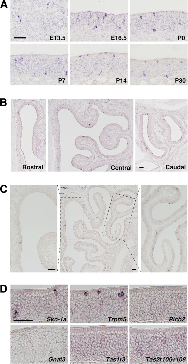
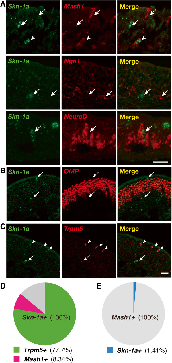
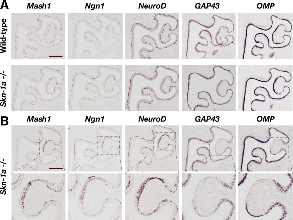
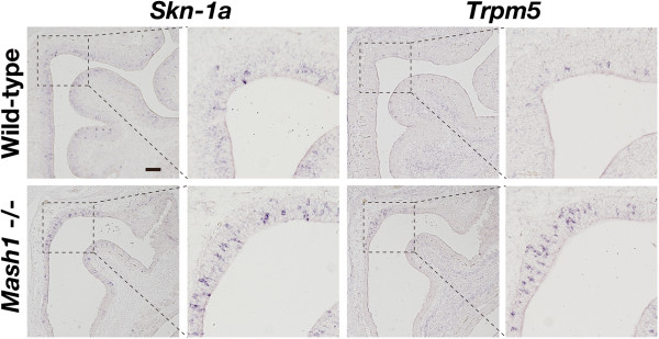
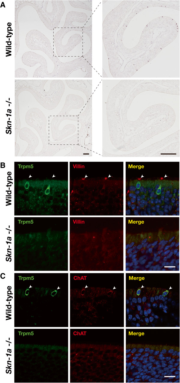
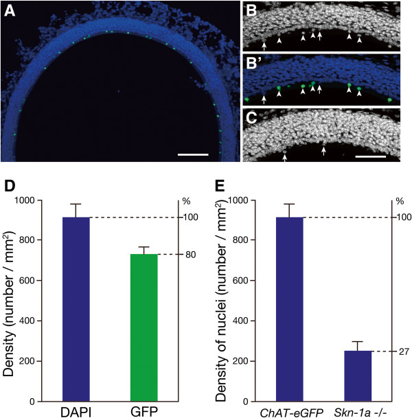
Results: Skn-1a is expressed in a subset of basal cells and apical non-neuronal cells in the MOE of embryonic and adult mice. Two-color in situ hybridization revealed that a small population of Skn-1a-expressing cells was co-labeled with Mash1/Ascl1 and that most Skn-1a-expressing cells coexpress Trpm5. To investigate whether Skn-1a has an irreplaceable role in the MOE, we analyzed Skn-1a-deficient mice. In the absence of Skn-1a, olfactory sensory neurons differentiate normally except for a limited defect in terminal differentiation in ectoturbinate 2 of some of MOEs examined. In contrast, the impact of Skn-1a deficiency on Trpm5-expressing microvillous cells is much more striking: Trpm5, villin, and choline acetyltransferase, cell markers previously shown to identify Trpm5-expressing microvillous cells, were no longer detectable in Skn-1a-deficient mice. In addition, quantitative analysis demonstrated that the density of superficial microvillous cells was significantly decreased in Skn-1a-deficient mice.
Conclusion: Skn-1a is expressed in a minority of Mash1-positive olfactory progenitor cells and a majority of Trpm5-expressing microvillous cells in the main olfactory epithelium. Loss-of-function mutation of Skn-1a resulted in complete loss of Trpm5-expressing microvillous cells, whereas most of olfactory sensory neurons differentiated normally. Thus, Skn-1a is a critical regulator for the generation of Trpm5-expressing microvillous cells in the main olfactory epithelium in mice.
Figures
Similar articles
-
Skn-1a/Pou2f3 functions as a master regulator to generate Trpm5-expressing chemosensory cells in mice.PLoS One. 2017 Dec 7;12(12):e0189340. doi: 10.1371/journal.pone.0189340. eCollection 2017. PLoS One. 2017. PMID: 29216297 Free PMC article.
-
Chemical Exposure-Induced Changes in the Expression of Neurotrophins and Their Receptors in the Main Olfactory System of Mice Lacking TRPM5-Expressing Microvillous Cells.Int J Mol Sci. 2018 Sep 27;19(10):2939. doi: 10.3390/ijms19102939. Int J Mol Sci. 2018. PMID: 30261693 Free PMC article.
-
Lack of TRPM5-Expressing Microvillous Cells in Mouse Main Olfactory Epithelium Leads to Impaired Odor-Evoked Responses and Olfactory-Guided Behavior in a Challenging Chemical Environment.eNeuro. 2017 Jun 12;4(3):ENEURO.0135-17.2017. doi: 10.1523/ENEURO.0135-17.2017. eCollection 2017 May-Jun. eNeuro. 2017. PMID: 28612045 Free PMC article.
-
TRPM5.Handb Exp Pharmacol. 2014;222:489-502. doi: 10.1007/978-3-642-54215-2_19. Handb Exp Pharmacol. 2014. PMID: 24756718 Review.
-
TRPM5 in the battle against diabetes and obesity.Acta Physiol (Oxf). 2018 Feb;222(2). doi: 10.1111/apha.12949. Epub 2017 Oct 4. Acta Physiol (Oxf). 2018. PMID: 28834354 Review.
Cited by
-
Thymic epithelial cell heterogeneity: TEC by TEC.Nat Rev Immunol. 2020 Apr;20(4):239-253. doi: 10.1038/s41577-019-0238-0. Epub 2019 Dec 5. Nat Rev Immunol. 2020. PMID: 31804611 Review.
-
OCA-T1 and OCA-T2 are coactivators of POU2F3 in the tuft cell lineage.Nature. 2022 Jul;607(7917):169-175. doi: 10.1038/s41586-022-04842-7. Epub 2022 May 16. Nature. 2022. PMID: 35576971 Free PMC article.
-
Dissecting the cellular specificity of smoking effects and reconstructing lineages in the human airway epithelium.Nat Commun. 2020 May 19;11(1):2485. doi: 10.1038/s41467-020-16239-z. Nat Commun. 2020. PMID: 32427931 Free PMC article.
-
Ascl3 transcription factor marks a distinct progenitor lineage for non-neuronal support cells in the olfactory epithelium.Sci Rep. 2016 Dec 2;6:38199. doi: 10.1038/srep38199. Sci Rep. 2016. PMID: 27910949 Free PMC article.
-
A nasal cell atlas reveals heterogeneity of tuft cells and their role in directing olfactory stem cell proliferation.Sci Immunol. 2024 Feb 2;9(92):eabq4341. doi: 10.1126/sciimmunol.abq4341. Epub 2024 Feb 2. Sci Immunol. 2024. PMID: 38306414 Free PMC article.
References
-
- Farbman A. Cell Biology of Olfactory Epithelium. 2. New York: Wiley-Liss; 2000.
Publication types
MeSH terms
Substances
Grants and funding
LinkOut - more resources
Full Text Sources
Other Literature Sources
Molecular Biology Databases
