. 2014 Jan 15;3(1):12-21.
doi: 10.1242/bio.20136205.

Functional coupling of chloride-proton exchanger ClC-5 to gastric H+,K+-ATPase

Affiliations

Functional coupling of chloride-proton exchanger ClC-5 to gastric H+,K+-ATPase

Yuji Takahashi et al. Biol Open. .

Abstract

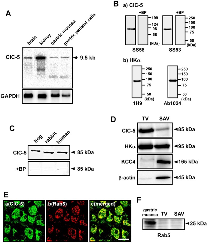
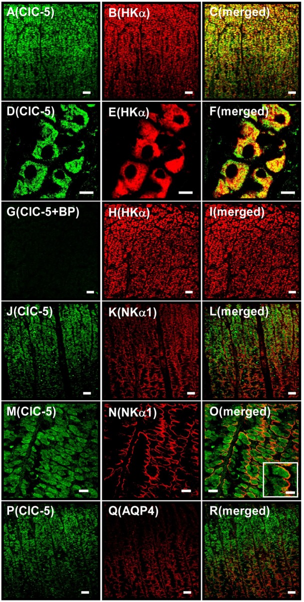
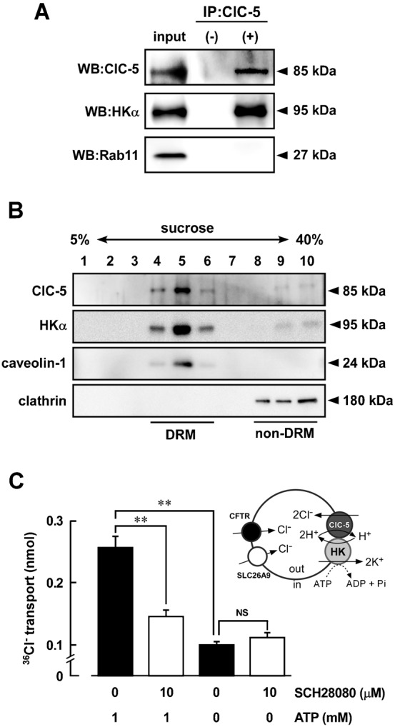
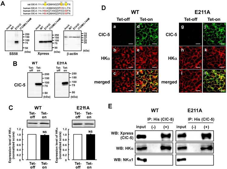
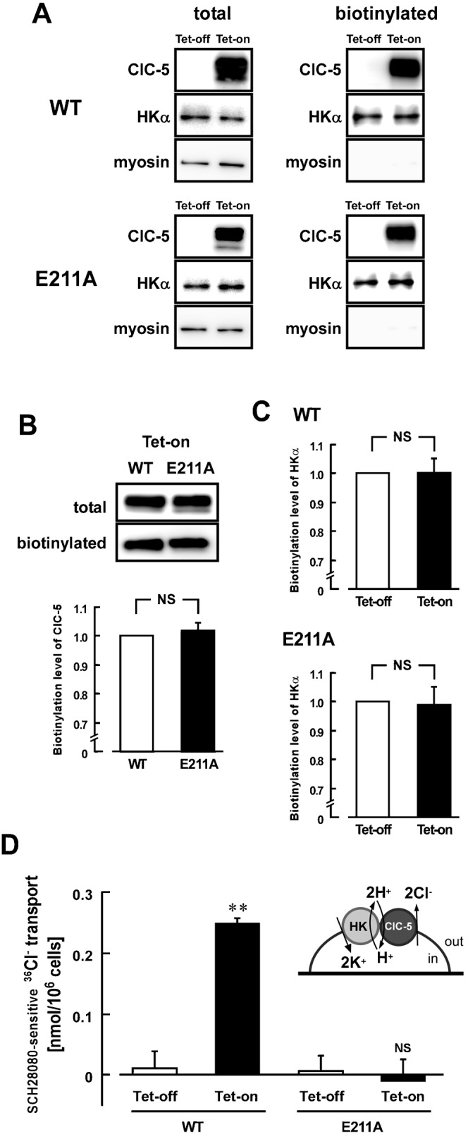
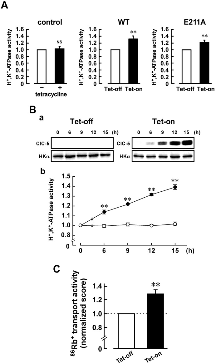
It has been reported that chloride-proton exchanger ClC-5 and vacuolar-type H(+)-ATPase are essential for endosomal acidification in the renal proximal cells. Here, we found that ClC-5 is expressed in the gastric parietal cells which secrete actively hydrochloric acid at the luminal region of the gland, and that it is partially localized in the intracellular tubulovesicles in which gastric H(+),K(+)-ATPase is abundantly expressed. ClC-5 was co-immunoprecipitated with H(+),K(+)-ATPase in the lysate of tubulovesicles. The ATP-dependent uptake of (36)Cl(-) into the vesicles was abolished by 2-methyl-8-(phenylmethoxy)imidazo[1,2-a]pyridine-3-acetonitrile (SCH28080), an inhibitor of H(+),K(+)-ATPase, suggesting functional expression of ClC-5. In the tetracycline-regulated expression system of ClC-5 in the HEK293 cells stably expressing gastric H(+),K(+)-ATPase, ClC-5 was co-immunoprecipitated with H(+),K(+)-ATPase, but not with endogenous Na(+),K(+)-ATPase. The SCH28080-sensitive (36)Cl(-) transporting activity was observed in the ClC-5-expressing cells, but not in the ClC-5-non-expressing cells. The mutant (E211A-ClC-5), which has no H(+) transport activity, did not show the SCH28080-sensitive (36)Cl(-) transport. On the other hand, both ClC-5 and its mutant (E211A) significantly increased the activity of H(+),K(+)-ATPase. Our results suggest that ClC-5 and H(+),K(+)-ATPase are functionally associated and that they may contribute to gastric acid secretion.

Keywords: ClC-5; Gastric acid; H+,K+-ATPase; Parietal cell; Tubulovesicle.


Conflict of interest statement

Competing interests: The authors have no competing interests to declare.

Figures

Fig. 1.
Fig. 1.. Expression of ClC-5 in gastric samples.
(A) Expression of ClC-5 mRNA in the stomach. Northern blotting was performed with poly A+ RNA (2.5 µg/lane) from brain, kidney and stomach of rabbits. A single band of 9.5 kb was detected with the ClC-5 cDNA probe. As a control, expression of GAPDH (1.3 kb) was examined. (B) Specificity of anti-ClC-5 and anti-H+,K+-ATPase α-subunit (HKα) antibodies. Western blotting was performed with hog gastric tubulovesicles (10 µg of protein) by using anti-ClC-5 antibodies (SS58 and SS53) (a) and anti-HKα antibodies (1H9 and Ab1024) (b). A single band of 85 kDa (a) or 95 kDa (b) was detected. The 85-kDa band was disappeared when the ClC-5 antibodies were preincubated with the corresponding blocking peptide (antibody:peptide  =  1:5) (+BP; a). (C) Western blotting was performed with hog and human gastric tubulovesicles and rabbit gastric P3 fraction (5, 50 and 40 µg of protein, respectively) by using anti-ClC-5 antibody (SS58). In each sample, an 85 kDa-band was detected (upper panel), and the band disappeared in the presence of the corresponding blocking peptide (lower panel). (D) Western blotting was performed with hog tubulovesicles (TV) and stimulation-associated vesicles (SAV) (10 µg of protein) by using anti-ClC-5 (SS58), anti-HKα (1H9), anti-KCC4 and anti-β-actin antibodies. ClC-5 (85 kDa) was predominantly expressed in TV, while KCC4 (165 kDa) and β-actin (45 kDa) were predominantly in SAV. HKα (95 kDa) was found in both TV and SAV. (E) a–c show the same tissue under a microscope. Double immunostaining was performed with hog gastric mucosa by using anti-ClC-5 (SS53) plus anti-Rab5 antibodies. Original magnification: ×63. Scale bars, 20 µm. (F) Western blotting was performed with TV, SAV and membrane fraction of gastric mucosa of hogs (30 µg of protein) by using anti-Rab5 antibody. Rab5 (25 kDa) was expressed in the gastric mucosa.
Fig. 2.
Fig. 2.. Immunostaining for ClC-5 in isolated hog gastric mucosa.
A–C show the same tissue under a microscope (as do D–F, G–I, J–L, M–O and P–R). Double immunostaining was performed with hog gastric mucosa by using anti-ClC-5 (SS53) plus anti-HKα (1H9) antibodies (A–I), anti-ClC-5 plus anti-NKα1 antibodies (J–O), and anti-ClC-5 plus anti-AQP4 antibodies (P–R). (A–F) Localizations of ClC-5 (A and D), HKα (B and E) and ClC-5 plus HKα (merged image; C and F) are shown. (G–I) Anti-ClC-5 antibody was pretreated with the blocking peptide. Localizations of ClC-5 (G), HKα (H) and ClC-5 plus HKα (merged image; I) were shown. Positive ClC-5 staining disappeared (G). (J–O) Localizations of ClC-5 (J and M), NKα1 (K and N) and ClC-5 plus NKα1 (merged image; L and O). In the inset of O, an enlarged image of a parietal cell is shown. (P–R) Localizations of ClC-5 (P), AQP4 (Q) and ClC-5 plus AQP4 (merged image; R). Original magnification: ×20 (A–C, G–L and P–R), ×40 (M–O) and ×63 (D–F). Scale bars, 50 µm (A–C, G–L and P–R), 10 µm (D–F and inset of O), and 20 µm (M–O).
Fig. 3.
Fig. 3.. Association of ClC-5 with H+,K+-ATPase in hog gastric tubulovesicles.
(A) Immunoprecipitation (IP) was performed with the detergent extracts of the hog gastric tubulovesicles (100 µg of potein) using anti-ClC-5 antibody (SS58) and protein A/G-agarose (IP: ClC-5, +). In control experiments, preimmune serum instead of the antibody was used (IP: ClC-5, −). The detergent extracts and immunoprecipitation samples were detected by Western blotting (WB) using antibodies for ClC-5 (SS58) labeled with HRP, HKα (1H9) and Rab11. The immunoprecipitation shown is representative of three independent experiments. (B) Detergent-resistant membrane (DRM) fractions and non-DRM fractions were isolated from hog gastric tubulovesicles by sucrose gradient (5–40%) as described in Materials and Methods. Western blotting was performed by using antibodies for ClC-5 (SS53), HKα (Ab1024), caveolin-1 and clathrin. (C) Inhibition of 36Cl uptake into tubulovesicles by an inhibitor of H+,K+-ATPase (SCH28080). The 36Cl uptake in tubulovesicles was measured as described in Materials and Methods. Effects of 1 mM ATP and/or 10 µM SCH28080 on the 36Cl uptake were examined. As a control, the uptake was measured in the absence of ATP. n = 10. NS, not significantly different (P>0.05); **, significantly different (P<0.01).
Fig. 4.
Fig. 4.. Tetracycline-regulated expression of ClC-5 in the HEK293 cells stably expressing gastric H+,K+-ATPase.
(A) Alignments of rat ClC-5, human ClC-5, human ClC-3 and human ClC-4 around an epitope of the anti-ClC-5 antibody are shown (upper panel). WT-ClC-5, E741D-ClC-5 and I732M/L744M-ClC-5 were transiently transfected in the HEK293 cells. In lower panels, Western blotting was performed with the membrane fraction (50 µg of protein) using anti-ClC-5 (SS58) (left), anti-Xpress (middle) and anti-β-actin (right) antibodies. No significant signal was observed in mock-transfected cells. (B) The tetracycline-regulated expression systems of WT-ClC-5 and E211A-ClC-5 were introduced to the HEK293 cells stably expressing H+,K+-ATPase. The cells were treated with (Tet-on) or without (Tet-off) 2 µg/ml tetracycline. Expression of WT- and E211A-ClC-5 in the membrane fraction of the cells (30 µg of protein) was confirmed by Western blotting using anti-Xpress antibody. (C) Expression level of HKα in the Tet-on cells was compared with that in the Tet-off cells. In the upper panel, a representative picture of Western blotting is shown. In the lower panel, the quantified score for the Tet-off cells is normalized as 1. n = 6. NS, P>0.05. (D) a–c show the same cells under a microscope (as do d–f, g–i, j–l). Double immunostaining was performed with the WT Tet-off cells (a–c), WT Tet-on cells (d–f), E211A Tet-off cells (g–i) and E211A Tet-on cells (j–l) using anti-Xpress (for ClC-5) plus anti-HKα (Ab1024) antibodies. Localizations of WT-ClC-5 (a and d), E211A-ClC-5 (g and j) and HKα (b, e, h and k), WT-ClC-5 plus HKα (merged images; c and f), and E211A-ClC-5 plus HKα (merged images; i and l) are shown. Scale bars, 20 µm. (E) WT-ClC-5 (left) and E211A-ClC-5 (right) are assembled to HKα in the HEK293 cells. Immunoprecipitation was performed with the detergent extracts of the Tet-on cells by using anti-His-tag antibody (for ClC-5) and protein A-agarose. The detergent extract (input) and the immunoprecipitation samples obtained with (IP: His(ClC-5), +) and without (IP: His(ClC-5), −) the antibody were detected by Western blotting (WB) using anti-Xpress antibody for detecting ClC-5 (top panel) and anti-HKα antibody (1H9; middle panel) and anti-NKα1 antibody (bottom panel; 100 kDa). In WB, anti-Xpress and anti-HKα antibodies were labeled with horseradish peroxidase. The immunoprecipitation shown is representative of three independent experiments.
Fig. 5.
Fig. 5.. Association of ClC-5 with gastric H+,K+-ATPase in the HEK293 cells.
(A) Cell surface biotinylation was performed with the Tet-on and Tet-off cells of WT-ClC-5 (upper panels) and E211A-ClC-5 (lower panels) prepared as described in Fig. 4. For detecting WT-ClC-5 (upper) or E211A-ClC-5 (lower), HKα and myosin (230 kDa) in the total lysate (left) and the biotinylated fraction (right), anti-Xpress (for ClC-5), anti-HKα (1H9) and anti-myosin antibodies were used, respectively. (B) Amount of biotinylated ClC-5 in the WT Tet-on cells is compared with that of biotinylated E211A-ClC-5 in the E211A Tet-on cells. In the upper panel, representative pictures of Western blotting are shown. In the lower panel, the biotinylated level was estimated by using the following equation: Calibrated biotinylation level of WT-ClC-5 (WT) or E211A-ClC-5 (E211A)  =  (amount of WT or E211A protein in the biotinylated fraction)/(amount of WT or E211A protein in the total lysate). The score for the WT cells is normalized as 1. n = 6. NS, P>0.05. (C) Amount of biotinylated HKα in the Tet-on cells is compared with that of the Tet-off cells (upper, WT; lower, E211A). The quantified score for the Tet-off cells is normalized as 1. n = 6. NS, P>0.05. (D) H+,K+-ATPase-dependent 36Cl transporting activity in the cells. 36Cl was loaded into the Tet-on and Tet-off cells of WT-ClC-5 and E211A-ClC-5 as described in Materials and Methods. The 36Cl transport activity was measured in the presence and absence of 10 µM SCH28080, and the SCH28080-sensitive (H+,K+-ATPase-dependent) transport activity was calculated. Significant activity was observed only in the WT Tet-on cells. n = 4–6. NS, not significantly different (P>0.05); **, significantly different (P<0.01).
Fig. 6.
Fig. 6.. Upregulation of the H+,K+-ATPase activity in the ClC-5-expressing cells.
(A) H+,K+-ATPase activity in the control HEK293 cells (left panels), and Tet-on and Tet-off cells of WT-ClC-5 (middle panels) and E211A-ClC-5 (right panels). The cells were treated with 2 µg/ml tetracycline for 12 h to obtain the Tet-on cells. The activity in the Tet-on cells was estimated by using the following equation: Calibrated H+,K+-ATPase activity = [(H+,K+-ATPase activity in the Tet-on cells)/(level of protein expression of HKα in the Tet-on cells)]/[(H+,K+-ATPase activity in the Tet-off cells)/(level of protein expression of HKα in the Tet-off cells)]. The H+,K+-ATPase activity of the WT and E211A Tet-off cells was 0.21±0.03 and 0.24±0.02 µmol Pi/mg of protein/h, respectively (n = 6). Calibrated H+,K+-ATPase activity of the Tet-off cells is normalized as 1. n = 6. **, P<0.01. (B) The upregulatory effect depends on the expression level of ClC-5 protein. The WT cells were treated with (Tet-on) and without (Tet-off) 2 µg/ml tetracycline for various period (6, 9, 12 and 15 h). In a, expression levels of ClC-5 and HKα proteins in the Tet-on (right) and Tet-off (left) cells are shown. In b, the H+,K+-ATPase activity of each sample was estimated as in Fig. 6A, and the data were shown as means ± s.e.m. (•, Tet-on; ○, Tet-off). n = 6. **, significantly different (P<0.01) compared with Tet-off. (C) 86Rb+ transport activity of the WT Tet-on and Tet-off cells. The 86Rb+ transport activity was measured in the presence and absence of 50 µM SCH28080, and the SCH28080-sensitive (H+,K+-ATPase-dependent) transport activity was calculated. The score was calculated by using the following equation: Normalized 86Rb+ transport activity = (the activity in the Tet-on cells)/(the activity in the Tet-off cells). The 86Rb+ transport activity of the Tet-off cells was 0.34±0.07 nmol 86Rb+/min/106 cells (n = 5). The score for Tet-off cells is normalized as 1. n = 5. **, P<0.01.

References

    1. Asano S., Inoie M., Takeguchi N. (1987). The Cl− channel in hog gastric vesicles is part of the function of H,K-ATPase. J. Biol. Chem. 262, 13263–13268. - PubMed
    1. Asano S., Arakawa S., Hirasawa M., Sakai H., Ohta M., Ohta K., Takeguchi N. (1994). C-terminal topology of gastric H+,K+-ATPase. Biochem. J. 299, 59–64. - PMC - PubMed
    1. Bamberg K., Nylander S., Helander K. G., Lundberg L. G., Sachs G., Helander H. F. (1994). In situ hybridization of mRNA for the gastric H+,K+-ATPase in rat oxyntic mucosa. Biochim. Biophys. Acta 1190, 355–359 10.1016/0005-2736(94)90094-9 - DOI - PubMed
    1. Calhoun B. C., Goldenring J. R. (1997). Two Rab proteins, vesicle-associated membrane protein 2 (VAMP-2) and secretory carrier membrane proteins (SCAMPs), are present on immunoisolated parietal cell tubulovesicles. Biochem. J. 325, 559–564. - PMC - PubMed
    1. Carmosino M., Procino G., Nicchia G. P., Mannucci R., Verbavatz J.-M., Gobin R., Svelto M., Valenti G. (2001). Histamine treatment induces rearrangements of orthogonal arrays of particles (OAPs) in human AQP4-expressing gastric cells. J. Cell Biol. 154, 1235–1244 10.1083/jcb.200103010 - DOI - PMC - PubMed

LinkOut - more resources