. 2014 Mar 20;123(12):1917-26.
doi: 10.1182/blood-2013-06-510180. Epub 2014 Jan 15.

Molecular mechanisms underlying synergistic adhesion of sickle red blood cells by hypoxia and low nitric oxide bioavailability

Affiliations

Molecular mechanisms underlying synergistic adhesion of sickle red blood cells by hypoxia and low nitric oxide bioavailability

Diana R Gutsaeva et al. Blood. .

Abstract

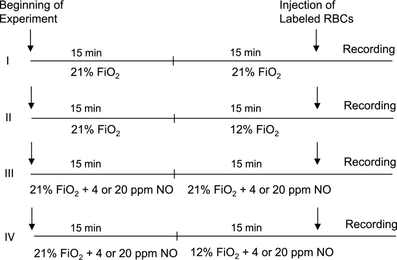
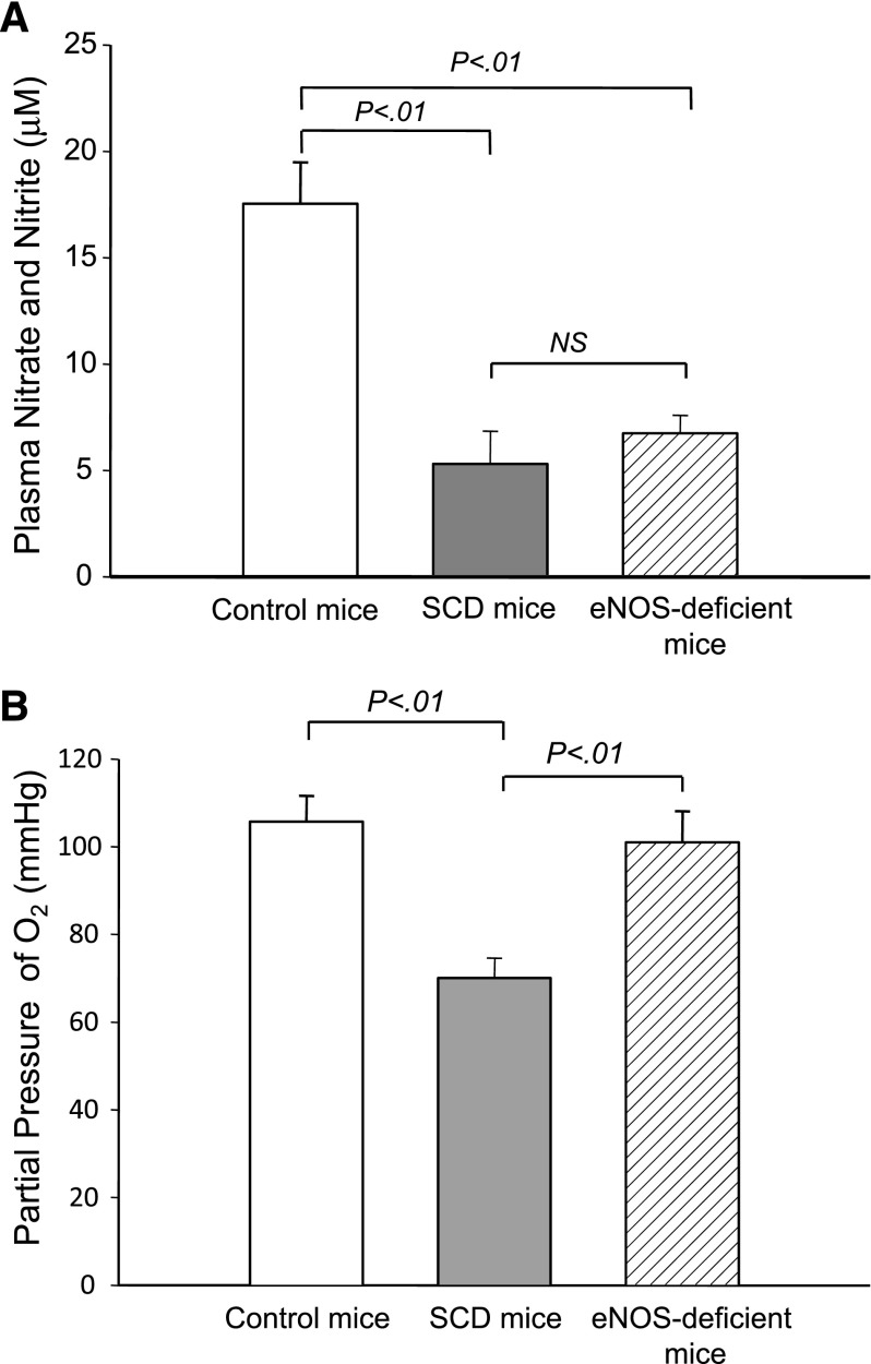
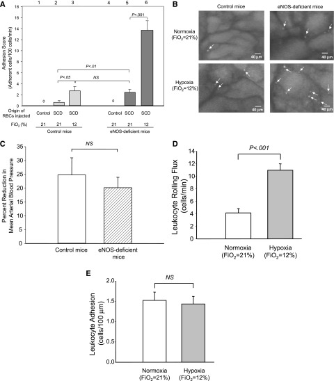
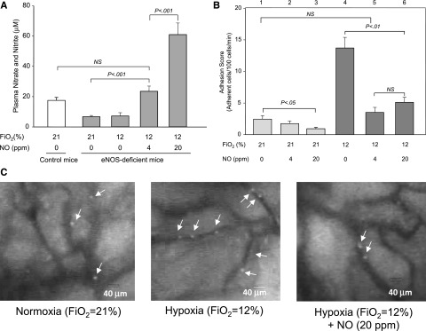
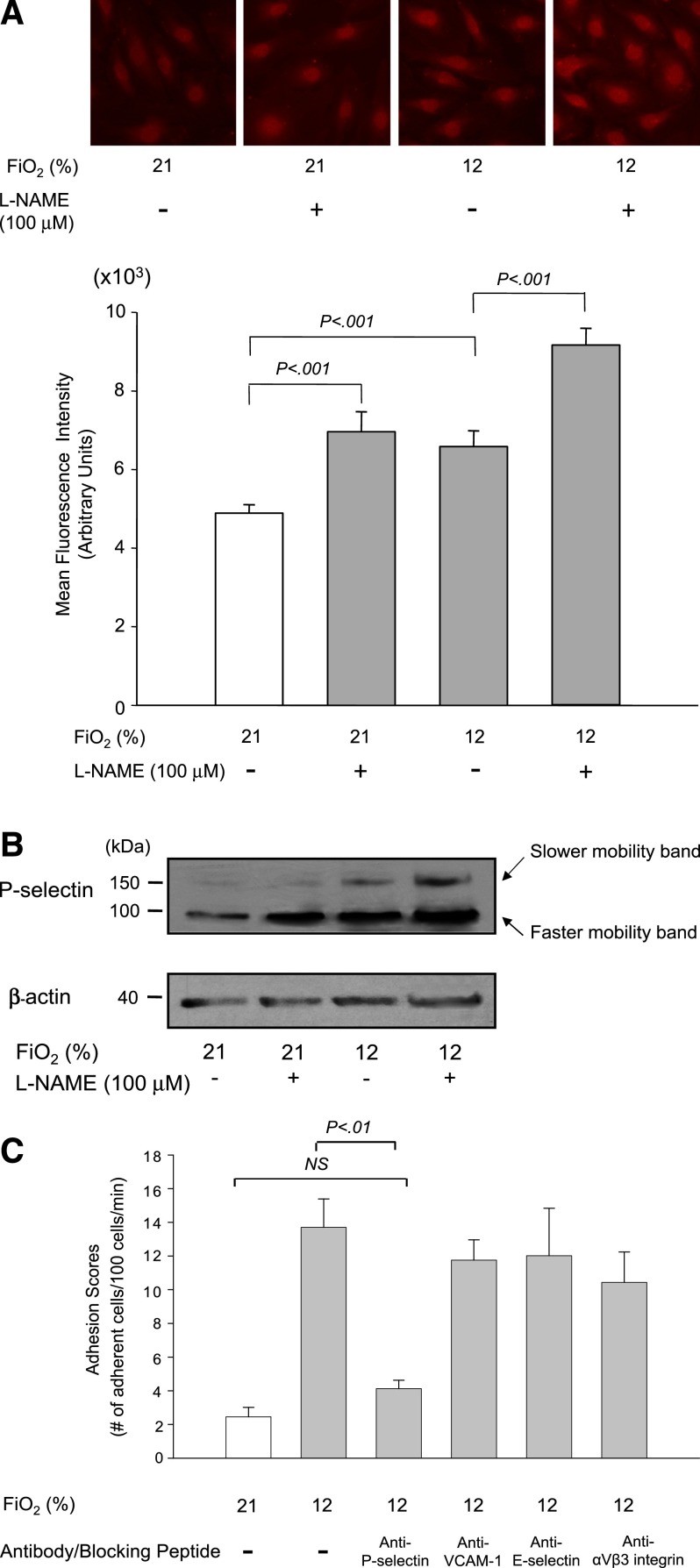
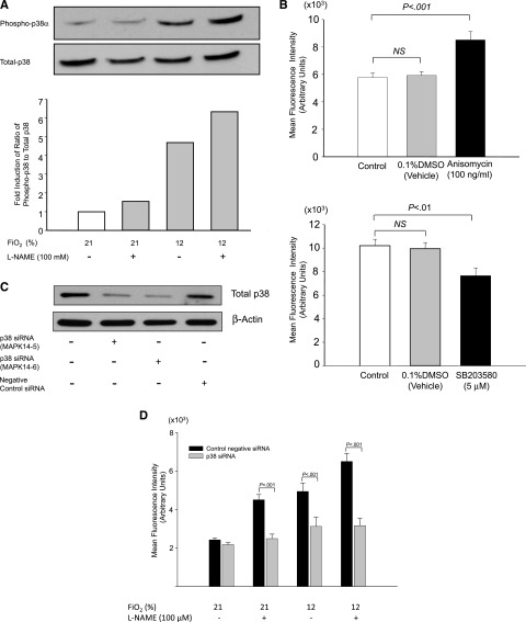
The molecular mechanisms by which nitric oxide (NO) bioavailability modulates the clinical expression of sickle cell disease (SCD) remain elusive. We investigated the effect of hypoxia and NO bioavailability on sickle red blood cell (sRBC) adhesion using mice deficient for endothelial NO synthase (eNOS) because their NO metabolite levels are similar to those of SCD mice but without hypoxemia. Whereas sRBC adhesion to endothelial cells in eNOS-deficient mice was synergistically upregulated at the onset of hypoxia, leukocyte adhesion was unaffected. Restoring NO metabolite levels to physiological levels markedly reduced sRBC adhesion to levels seen under normoxia. These results indicate that sRBC adherence to endothelial cells increases in response to hypoxia prior to leukocyte adherence, and that low NO bioavailability synergistically upregulates sRBC adhesion under hypoxia. Although multiple adhesion molecules mediate sRBC adhesion, we found a central role for P-selectin in sRBC adhesion. Hypoxia and low NO bioavailability upregulated P-selectin expression in endothelial cells in an additive manner through p38 kinase pathways. These results demonstrate novel cellular and signaling mechanisms that regulate sRBC adhesion under hypoxia and low NO bioavailability. Importantly, these findings point us toward new molecular targets to inhibit cell adhesion in SCD.


Figures

Figure 1
Figure 1
Treatment protocol of mice to study the effects of hypoxia and NO bioavailability on sRBC adhesion. Control and eNOS-deficient mice were treated with 4 different procedures (I-IV). The numbers of mice used for the treatments were as follows: I, 5 control mice and 5 eNOS-deficient mice; II, 5 control mice and 7 eNOS-deficient mice; III, 7 eNOS-deficient mice; and IV, 4 eNOS-deficient mice. A bolus of 2,7-bis-(carboxyethyl)-5-(and-6) carboxyfluorescein (BCECF)-labeled RBCs was injected into the mice, and 3 minutes later, the recording of microcirculation images was initiated and completed by analyzing 4 1-minute video segments. Fio2 indicates fraction of inspired oxygen in a gas mixture.
Figure 2
Figure 2
NO metabolite levels and Pao2 in control and eNOS-deficient mice. (A) Steady-state plasma nitrate/nitrite levels in control, SCD, and eNOS-deficient mice. Plasma nitrate/nitrite levels were determined using the Nitrate/Nitrite Colorimetric Assay Kit (Cayman Chemical). NS, not significant. (B) Pao2 levels in control, SCD, and eNOS-deficient mice. Arterial blood was drawn at baseline conditions for blood gas analysis. Values were mean ± SEM obtained from 3 to 4 mice in each group. P values are shown at the top of the figure.
Figure 3
Figure 3
Adhesion of sRBC and leukocytes in control and eNOS-deficient mice under normoxia (procedure I) or hypoxia (procedure II). (A) Analysis of RBC adhesion to endothelial cells by intravital microscopy. Labeled RBCs were prepared from either control or SCD mice and injected into control or eNOS-deficient mice. Note that RBCs prepared from control mice did not adhere to endothelial cells in either control or SCD mice, suggesting that the intravital microscopy analysis eliminates false-positive cell–cell interactions. Lanes are shown at the top of figure, and the origins of injected RBCs and oxygen tensions are shown at the bottom of figure. Values were mean ± SEM obtained from 3 to 7 mice in each group. P values are shown at the top of the figure. (B) Frame-captured images from videotaped intravital microscopy of bone marrow venules in control (left panels) and eNOS-deficient mice (right panels) after injection of BCECF-labeled sRBCs under normoxia (upper panels) and hypoxia (low panels). Arrows indicate adhered sRBCs. (C) Percent reduction in MABP in response to hypoxia (Fio2 = 12%) in control and eNOS-deficient mice. Values were mean ± SEM from 5 to 7 mice in each group. (D-E) Leukocyte rolling (D) and leukocyte adhesion (E) to endothelial cells in eNOS-deficient mice at normoxia (procedure I) or at hypoxia (procedure II). PE rat anti-CD45 antibody was injected into eNOS-deficient mice. Leukocyte adhesion was quantified by counting the number of adherent cells (stationary for >30 seconds) in a 100-μM length of the vessel. Note that only leukocyte rolling is increased at the onset of hypoxia. Values were mean ± SEM from 5 to 7 mice in each group. P values are shown in the figure.
Figure 4
Figure 4
Effects of NO inhalation on NO metabolite levels and sRBC adhesion in eNOS-deficient mice. (A) Plasma nitrate/nitrite levels in eNOS-deficient mice that inhaled NO gas under normoxia or hypoxia. Plasma nitrate/nitrite levels were determined using the Nitrate/Nitrite Colorimetric Assay Kit (Cayman Chemical). Values were mean ± SEM from 3 to 4 mice in each group. P values are shown in the figure. (B) Modulation by NO inhalation of sRBC adhesion in postcapillary venules of eNOS-deficient mice. eNOS-deficient mice were treated with 4 or 20 ppm NO gas at normoxia (procedure III) or hypoxia (procedure IV). Values were mean ± SEM from 5 to 7 mice in each group. P values are shown in the figure. (C) Frame-captured images from videotaped intravital microscopy of bone marrow postcapillary venules in eNOS-deficient mice after injection of BCECF-labeled sRBCs under normoxia, hypoxia, and hypoxia with 20 ppm NO. Arrows indicate adhered sRBCs.
Figure 5
Figure 5
Expression of P-selectin on HUVECs is regulated by hypoxia and NO bioavailability and its role in sRBC adhesion. (A) Effects of hypoxia and the eNOS inhibitor l-NAME on P-selectin expression on HUVECs. P-selectin expression on the cell surface of HUVECs was evaluated by immunofluorescence microscopy. Cells were treated with room air (Fio2 = 21%) or hypoxia of 12% O2 for 1 hour with or without l-NAME (100 μM). Images were taken at ×400 (upper panel). MFIs were calculated by scanning fluorescence images with ImageJ v1.43 (NIH). Results with MFI for each experiment are shown in the lower panel. Values were mean ± SEM from 3 to 4 images taken for each treatment with at least 5 cells analyzed per image. (B) Analysis of P-selectin expression in HUVECs by western blotting. Cells were treated as described previously, and the cell membrane fractions were isolated and subjected to western blot analysis. Note that the intensity of a slower-mobility protein band at 140 kDa is increased in response to hypoxia and l-NAME treatment. (C) Effect of blocking antibodies on sRBC adhesion in eNOS-deficient mice under hypoxia. eNOS-deficient mice were infused with antibody against P-selectin, VCAM-1, and E-selectin or αvβ3-integrin blocking peptide before intravital experiments, and the adhesion scores of sRBCs were measured. Values were mean ± SEM from 5 to 7 mice in each group. P values are shown at the top of the figure.
Figure 6
Figure 6
P-selectin expression in HUVECs is regulated by p38 kinase pathways. (A) Phosphorylation of p38 kinases increases in HUVECs treated with hypoxia for 1 hour with or without the NOS inhibitor l-NAME (100 μM). A representative blot was shown from 3 independent experiments. (B) P-selectin expression on the cell surface of HUVECs is regulated by p38 kinases. HUVECs were treated with anisomycin (100 ng/mL) (upper panel) or SB203580 (5 μM) (lower panel) for 1 hour, and P-selectin expression on the cell surface was evaluated by immunofluorescence microscopy as described in “Materials and methods.” MFIs were calculated by scanning fluorescence images with ImageJ v1.43 (NIH). (C) Knockdown of p38 kinase expression in HUVECs by siRNA constructs. Western blot showing that p38 siRNA, but not control negative siRNA, decreased total p38 protein by 50% to 66% in HUVECs at 48 hours. (D) P-selectin expression in HUVECs transfected with siRNA constructs for p38α kinase (gray columns) and negative control siRNA (black columns). Expression of P-selectin on the cell surface of HUVECs was quantitated by immunofluorescence microscopy as described in “Materials and methods,” and MFIs were calculated by scanning fluorescence images with ImageJ v1.43 (NIH).

Comment in

References

    1. Hebbel RP, Boogaerts MA, Eaton JW, Steinberg MH. Erythrocyte adherence to endothelium in sickle-cell anemia. A possible determinant of disease severity. N Engl J Med. 1980;302(18):992–995. - PubMed
    1. Ferrone FA, Hofrichter J, Eaton WA. Kinetics of sickle hemoglobin polymerization. I. Studies using temperature-jump and laser photolysis techniques. J Mol Biol. 1985;183(4):591–610. - PubMed
    1. Ataga KI, Smith WR, De Castro LM, et al. ICA-17043-05 Investigators. Efficacy and safety of the Gardos channel blocker, senicapoc (ICA-17043), in patients with sickle cell anemia. Blood. 2008;111(8):3991–3997. - PubMed
    1. Frenette PS. Sickle cell vaso-occlusion: multistep and multicellular paradigm. Curr Opin Hematol. 2002;9(2):101–106. - PubMed
    1. Morris CR, Kuypers FA, Larkin S, Vichinsky EP, Styles LA. Patterns of arginine and nitric oxide in patients with sickle cell disease with vaso-occlusive crisis and acute chest syndrome. J Pediatr Hematol Oncol. 2000;22(6):515–520. - PubMed

Publication types

MeSH terms