. 2014 Apr;88(7):3705-18.
doi: 10.1128/JVI.03039-13. Epub 2014 Jan 15.

Nod-like receptor X-1 is required for rhinovirus-induced barrier dysfunction in airway epithelial cells

Affiliations

Nod-like receptor X-1 is required for rhinovirus-induced barrier dysfunction in airway epithelial cells

Benjamin L Unger et al. J Virol. 2014 Apr.

Abstract

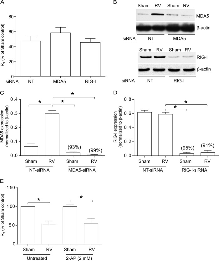
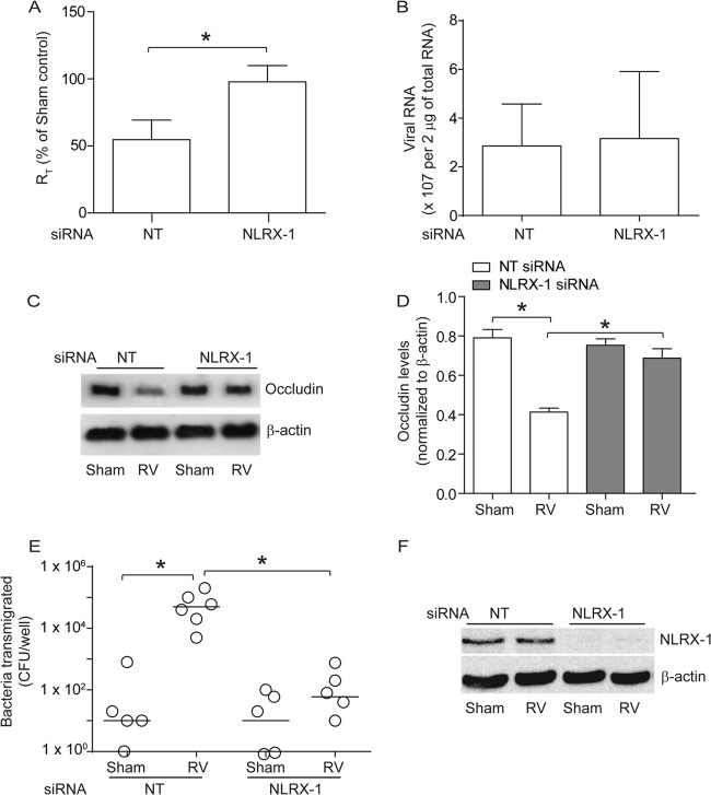
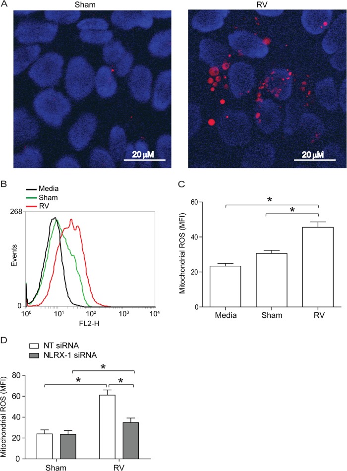
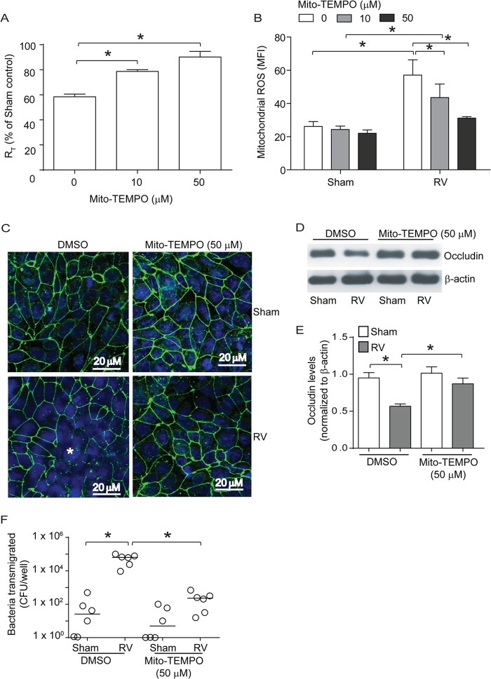
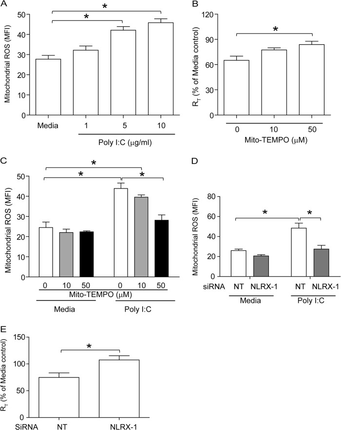
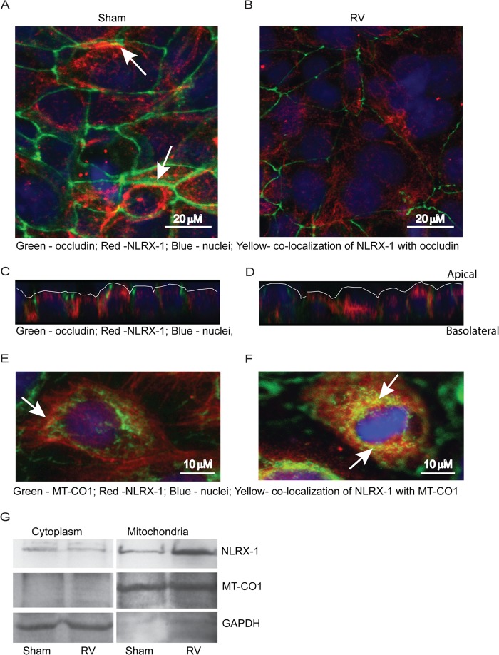
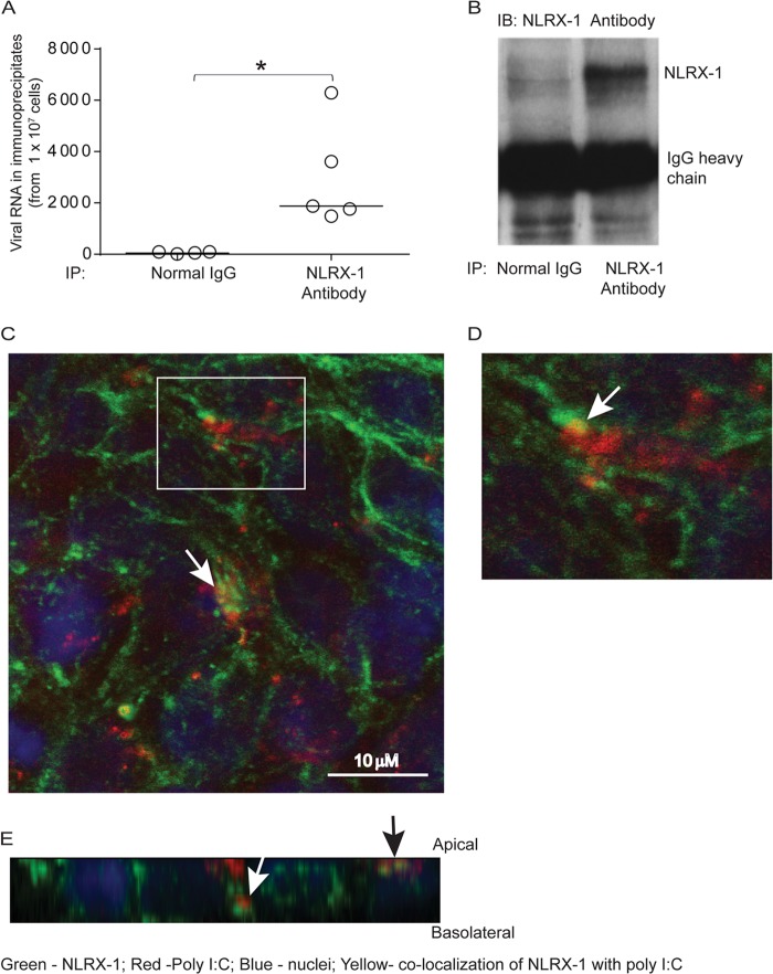
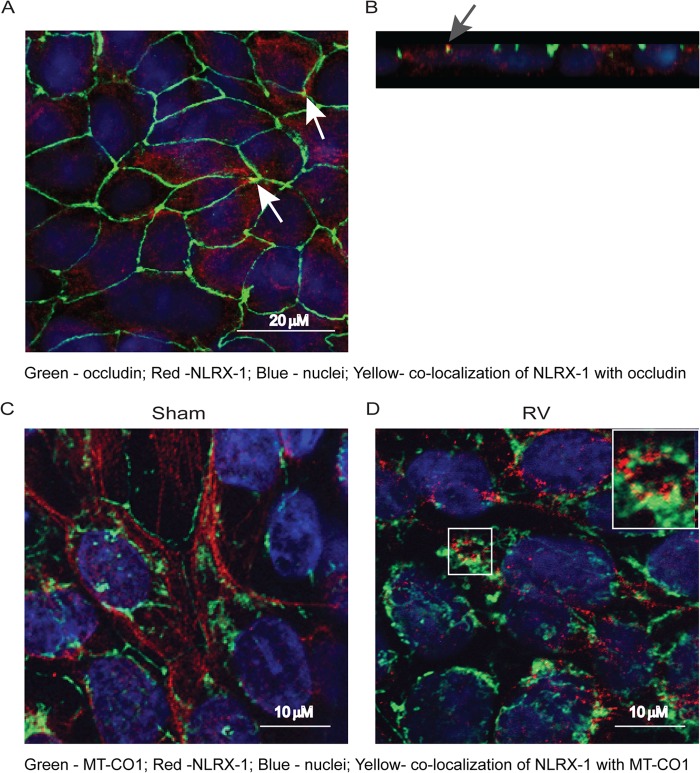
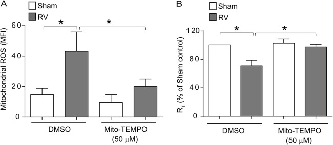
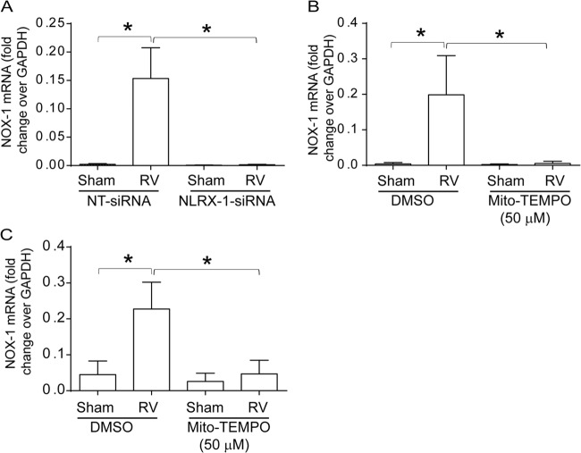
Barrier dysfunction of airway epithelium may increase the risk for acquiring secondary infections or allergen sensitization. Both rhinovirus (RV) and polyinosinic-polycytidilic acid [poly(I·C)], a double-stranded RNA (dsRNA) mimetic, cause airway epithelial barrier dysfunction, which is reactive oxygen species (ROS) dependent, implying that dsRNA generated during RV replication is sufficient for disrupting barrier function. We also demonstrated that RV or poly(I·C)-stimulated NADPH oxidase 1 (NOX-1) partially accounts for RV-induced ROS generation. In this study, we identified a dsRNA receptor(s) contributing to RV-induced maximal ROS generation and thus barrier disruption. We demonstrate that genetic silencing of the newly discovered dsRNA receptor Nod-like receptor X-1 (NLRX-1), but not other previously described dsRNA receptors, abrogated RV-induced ROS generation and reduction of transepithelial resistance (R(T)) in polarized airway epithelial cells. In addition, both RV and poly(I·C) stimulated mitochondrial ROS, the generation of which was dependent on NLRX-1. Treatment with Mito-Tempo, an antioxidant targeted to mitochondria, abolished RV-induced mitochondrial ROS generation, reduction in R(T), and bacterial transmigration. Furthermore, RV infection increased NLRX-1 localization to the mitochondria. Additionally, NLRX-1 interacts with RV RNA and poly(I·C) in polarized airway epithelial cells. Finally, we show that NLRX-1 is also required for RV-stimulated NOX-1 expression. These findings suggest a novel mechanism by which RV stimulates generation of ROS, which is required for disruption of airway epithelial barrier function.

Importance: Rhinovirus (RV), a virus responsible for a majority of common colds, disrupts the barrier function of the airway epithelium by increasing reactive oxygen species (ROS). Poly(I·C), a double-stranded RNA (dsRNA) mimetic, also causes ROS-dependent barrier disruption, implying that the dsRNA intermediate generated during RV replication is sufficient for this process. Here, we demonstrate that both RV RNA and poly(I·C) interact with NLRX-1 (a newly discovered dsRNA receptor) and stimulate mitochondrial ROS. We show for the first time that NLRX-1 is primarily expressed in the cytoplasm and at the apical surface rather than in the mitochondria and that NLRX-1 translocates to mitochondria following RV infection. Together, our results suggest a novel mechanism for RV-induced barrier disruption involving NLRX-1 and mitochondrial ROS. Although ROS is necessary for optimal viral clearance, if not neutralized efficiently, it may increase susceptibility to secondary infections and alter innate immune responses to subsequently inhaled pathogens, allergens, and other environmental factors.


Figures

FIG 1
FIG 1
Knockdown of MDA5 or RIG-I or inhibition of PKR does not affect RV-induced changes in RT. 16HBE14o− cells were transfected with NT, MDA5, or RIG-I siRNA, and the cells were allowed to polarize. (A) Cells were infected apically with RV or sham control, and RT was measured after 24 h. (B) Total proteins from cells transfected with NT, MDA5, or RIG-I siRNA were subjected to Western blot analysis with antibodies to MDA5, RIG-I, or β-actin. (C and D) Band intensities were quantified by NIH Image, and levels of MDA5 or RIG-I were normalized to the respective β-actin controls. (E) Cells were pretreated with 2-AP for 1 h, infected with RV or sham control, and incubated for 24 h in the presence or absence of 2-AP, and then RT was measured. The data in panels A, C, D, and E represent means and standard deviations (SD) calculated from 3 to 6 independent experiments (*, P ≤ 0.05; ANOVA). The percentages in panels C and D represent percent inhibition of MDA5 and RIG-I expression, respectively, in gene-specific siRNA-transfected cells compared to NT siRNA-transfected cells. The images in panel B are representative of 3 independent experiments.
FIG 2
FIG 2
NLRX-1 is required for RV-induced tight-junction breakdown. 16HBE14o− cells were transfected with NT or NLRX-1 siRNA and allowed to polarize for 48 h. (A) Cells were infected with either RV or sham control and incubated for 24 h, and then RT was measured. (B) Total RNA isolated from sham- or RV-infected cells was used to determine viral RNA copy numbers. (C and D) NP-40-insoluble fractions from cells infected with sham control or RV were subjected to Western blot analysis using antibodies to occludin or β-actin, and band intensities were quantified and expressed as fold change over β-actin. (E) NT or NLRX-1 siRNA-transfected cell cultures were infected with RV or sham control and incubated for 16 h. NTHI was added to the apical surface and incubated for an additional 4 h, and the number of bacteria in the basolateral chamber was determined by plating. (F) Total proteins from cells transfected with NT or NLRX-1 siRNA and infected with sham control or RV were subjected to Western blot analysis with antibody to NLRX-1 or β-actin to confirm knockdown of NLRX-1. The data in panels A, B, and D represent means and SD calculated from 3 or 4 independent experiments (*, P ≤ 0.05; ANOVA). The data in panel D represent medians with ranges from 3 independent experiments (*, P ≤ 0.05; ANOVA on ranks). The images in panels B and E are representatives of 3 or 4 independent experiments.
FIG 3
FIG 3
RV stimulates NLRX-1-dependent mitochondrial ROS in polarized airway epithelial cells. The polarized monolayers of 16HBE14o− cells were infected with RV or sham control and incubated for 90 min at 33°C. The infection medium was replaced with fresh medium, and incubation continued for an additional 16 h. The cells were washed once with HBSS and incubated with 5 μM MitoSox Red for 15 min at 37°C. The cells were washed with warm HBSS. (A) Cells were incubated with HBSS containing Hoechst dye (nuclear stain) for 10 min, which was then replaced with warm HBSS, and the cells were imaged immediately. The images are representative of 3 independent experiments (red, MitoSox Red fluorescence indicative of mitochondrial ROS; blue, nuclei). (B and C) Cells were detached from the plate, suspended in warm HBSS, and analyzed by flow cytometry. MFI, mean fluorescence intensity. (D) NT or NLRX-1 siRNA-transfected cells were infected with sham control or RV, and mitochondrial ROS was quantified by flow cytometry 16 h postinfection. (B) Representative histogram of 3 independent experiments. (C and D) Data represent means and SD calculated from 3 or 4 independent experiments (*, P ≤ 0.05; ANOVA).
FIG 4
FIG 4
Mito-Tempo, an antioxidant targeted to mitochondria, blocks RV-induced reduction in RT and mitochondrial ROS generation. Polarized monolayers of 16HBE14o− cells were treated with 0, 10, or 50 μM Mito-Tempo for 1 h both apically and basolaterally. (A and B) Cells were infected with RV or sham control apically and incubated for 90 min. The infection medium was replaced with fresh medium containing 0, 10, or 50 μM Mito-Tempo and incubated for an additional 24 h. The RT was measured, and then the cells were washed and incubated with MitoSox Red as described for Fig. 3 and analyzed by flow cytometry. (C) Polarized cells were infected with RV or sham control in the presence of 0 or 50 μM Mito-Tempo and incubated for 24 h. The cells were fixed, blocked with 1% BSA in PBS, and incubated with antibody to occludin. Bound antibody was detected with antirabbit IgG conjugated with Alexa Fluor 488, and the cells were counterstained with DAPI and then subjected to indirect immunofluorescence microscopy. The images are representative of 3 or 4 independent experiments (*, dissociation of occludin from the tight-junction complex; green, occludin; blue, nuclei). (D and E) NP-40-insoluble fractions from cells infected with RV or sham control in the presence or absence of Mito-Tempo were subjected to Western blot analysis, and the band intensities were quantified using NIH Image J and expressed as fold change over β-actin. (F) Serial dilutions of basolateral media from cell cultures infected with RV or sham control in the presence or absence of Mito-Tempo were plated to determine the number of bacteria transmigrated from the apical to the basolateral surface. The data in panels A, B, and D represent means and SD calculated from 3 or 4 independent experiments (*, P ≤ 0.05; ANOVA). The data in panel F represent medians with ranges from 3 independent experiments (*, P ≤ 0.05; ANOVA on ranks).
FIG 5
FIG 5
Poly(I·C) induces mitochondrial ROS in polarized 16HBE14o− cells in an NLRX-1- dependent manner. (A) Cells were treated with 1, 5, or 10 μg/ml poly(I·C) and incubated for 6 h at 37°C. Mitochondrial ROS was determined by flow cytometry as described for Fig. 3. (B and C) Polarized monolayers of 16HBE14o− cells were incubated with 0, 10, or 50 μM Mito-Tempo for 1 h both apically and basolaterally and then treated with 5 μg/ml poly(I·C) in the presence of 0, 10, or 50 μM Mito-Tempo. After 6 h, RT was measured, and then the cells were treated with MitoSox Red to determine the levels of mitochondrial ROS. (D and E) Cells were transfected with NT or NLRX-1 siRNA, allowed to polarize for 2 days, and then treated with 5 μg/ml poly(I·C). After 6 h, RT was measured, and then the cells were treated with MitoSox Red to determine the levels of mitochondrial ROS. The data represent means and SD calculated from 3 independent experiments (*, P ≤ 0.05; ANOVA).
FIG 6
FIG 6
NLRX-1 redistributes in airway epithelial cells following RV infection. (A to F) Polarized 16HBE14o− cells were infected with RV or sham control. After 16 h, the cells were fixed and incubated with a mixture of antibodies to occludin and NLRX-1 (A to D) or a mixture of antibodies to MT-CO1 (mitochondrial protein) and NLRX-1 (E and F). Bound antibodies were detected by secondary antibodies conjugated with either Alexa Fluor 598 (NLRX-1) or Alexa Fluor 488 (occludin [A to D] and MT-CO1 [E and F]). The cells were counterstained with DAPI and subjected to indirect immunofluorescence microscopy. The arrows in panel A represent colocalization (yellow) of NLRX-1 with occludin. Panels C and D are the Z sections of panels A and B, respectively, showing the distribution of NLRX-1 in relation to occludin, and the apical surface of the cultures is marked by the white line. The arrows in panels E and F represent colocalization (yellow) of NLRX-1 with MT-CO1. (G) Cytoplasmic or mitochondrial proteins isolated from sham- or RV-infected cells were subjected to Western blot analysis with antibodies to NLRX-1, MT-CO1, or GAPDH. The absence of a band corresponding to MT-CO1 in the cytoplasmic fraction and the absence of GAPDH in the mitochondrial fraction indicate efficient separation of cytoplasmic proteins from mitochondrial proteins. The images are representative of 3 or 4 experiments.
FIG 7
FIG 7
NLRX-1 interacts with viral RNA and poly(I·C). The polarized airway epithelial cells were RV or sham infected and incubated for 16 h, and cell lysates were immunoprecipitated with normal IgG or anti-NLRX-1 antibody. (A and B) Total RNA was isolated from the immunoprecipitates, the RV RNA copy number was determined (A), and an aliquot of immunoprecipitates (IP) was subjected to Western blot (IB) analysis with NLRX-1 antibody (B). (C to E) Rhodamine-labeled poly(I·C) was added to the apical surfaces of polarized airway epithelial cells and incubated for 3 h. The cells were fixed in methanol, blocked with BSA, incubated with antibody to NLRX-1 conjugated with Alexa Fluor 488, and counterstained with DAPI. The cells were visualized by confocal microscopy. Panel D is a magnified view of the boxed area in panel C. Panel E shows a Z section of panel C. The white arrows in panels C and D indicate colocalization of poly(I·C) with NLRX-1. The white and black arrows in panel E represent NLRX-1 colocalization with poly(I·C) in the subapical and apical locations, respectively. The data in panel A represent medians and ranges from 5 independent experiments (*, P ≤ 0.05; ANOVA). The images are representative of three or five independent experiments.
FIG 8
FIG 8
RV promotes NLRX-1 translocation to mitochondria in primary airway epithelial cells. (A and B) Mucociliary differentiated primary airway epithelial cells were fixed and immunolabeled with a mixture of antibodies to occludin (green) and NLRX-1 (red). Panel B is a Z section of panel A showing NLRX-1 on the apical surface and in the cytoplasm. The arrows indicate colocalization of NLRX-1 with occludin. (C and D) Mucociliary differentiated primary airway epithelial cells were apically infected with sham control or RV, incubated for 24 h, fixed, immunolabeled with antibodies to NLRX-1 (red) and MT-CO1 (green), counterstained with DAPI, and subjected to confocal microscopy. The images represent optical sections taken at a depth of 9 μm to show both NLRX-1 and MT-CO1, presumably in the middle of the cells. The total thickness of cell cultures was approximately 20 μm. The inset in panel D is a magnified view of the boxed area. All images are representative of three independent experiments.
FIG 9
FIG 9
RV-stimulated mitochondrial ROS is required for RT reduction in primary airway epithelial cells. Mucociliary differentiated primary airway epithelial cells were apically infected with sham control or RV in the presence or absence of 50 μM Mito-Tempo and incubated for 24 h. (A) Cells were incubated with MitoSox Red and analyzed by flow cytometry to determine the levels of mitochondrial ROS. (B) RT was measured and expressed as a percentage of sham-infected vehicle-treated controls. The data represent means and SD calculated from three independent experiments (*, P ≤ 0.05; ANOVA).
FIG 10
FIG 10
Inhibition of NLRX-1 or mitochondrial ROS blocks RV-induced NOX-1 expression. (A) 16HBE14o− cells were transfected with NT or NLRX-1 siRNA, infected with sham control or RV, and incubated for 16 h. (B and C) 16HBE14o− cells or mucociliary differentiated cells were infected with RV or sham control in the presence or absence of Mito-Tempo and incubated for 16 h. Total RNA was harvested, and the expression of NOX-1 mRNA was determined by qPCR and expressed as the fold change over the GAPDH housekeeping gene. The data represent means and SD calculated from 3 independent experiments (*, P ≤ 0.05; ANOVA).

Similar articles

Cited by

References

    1. Coyne CB, Bergelson JM. 2006. Virus-induced Abl and Fyn kinase signals permit coxsackievirus entry through epithelial tight junctions. Cell 124:119–131. 10.1016/j.cell.2005.10.035 - DOI - PubMed
    1. Bergelson JM, Cunningham JA, Droguett G, Kurt-Jones EA, Krithivas A, Hong JS, Horwitz MS, Crowell RL, Finberg RW. 1997. Isolation of a common receptor for Coxsackie B viruses and adenoviruses 2 and 5. Science 275:1320–1323. 10.1126/science.275.5304.1320 - DOI - PubMed
    1. Evans MJ, von Hahn T, Tscherne DM, Syder AJ, Panis M, Wolk B, Hatziioannou T, McKeating JA, Bieniasz PD, Rice CM. 2007. Claudin-1 is a hepatitis C virus co-receptor required for a late step in entry. Nature 446:801–805. 10.1038/nature05654 - DOI - PubMed
    1. Zheng A, Yuan F, Li Y, Zhu F, Hou P, Li J, Song X, Ding M, Deng H. 2007. Claudin-6 and claudin-9 function as additional coreceptors for hepatitis C virus. J. Virol. 81:12465–12471. 10.1128/JVI.01457-07 - DOI - PMC - PubMed
    1. Antar AA, Konopka JL, Campbell JA, Henry RA, Perdigoto AL, Carter BD, Pozzi A, Abel TW, Dermody TS. 2009. Junctional adhesion molecule-A is required for hematogenous dissemination of reovirus. Cell Host Microbe 5:59–71. 10.1016/j.chom.2008.12.001 - DOI - PMC - PubMed

Publication types

LinkOut - more resources