. 2014 Jan;6(1):70-81.
doi: 10.18632/aging.100629.

Serines 440 and 467 in the Werner syndrome protein are phosphorylated by DNA-PK and affects its dynamics in response to DNA double strand breaks

Affiliations

Serines 440 and 467 in the Werner syndrome protein are phosphorylated by DNA-PK and affects its dynamics in response to DNA double strand breaks

Rika Kusumoto-Matsuo et al. Aging (Albany NY). 2014 Jan.

Abstract

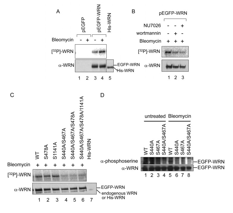
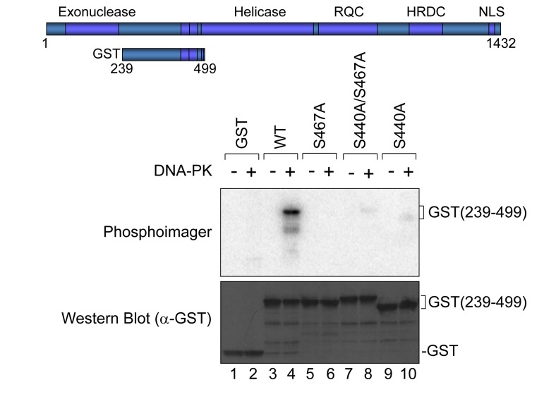
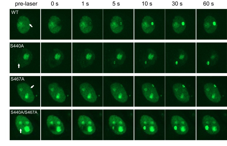
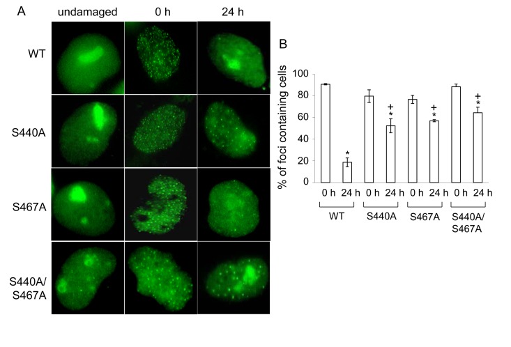
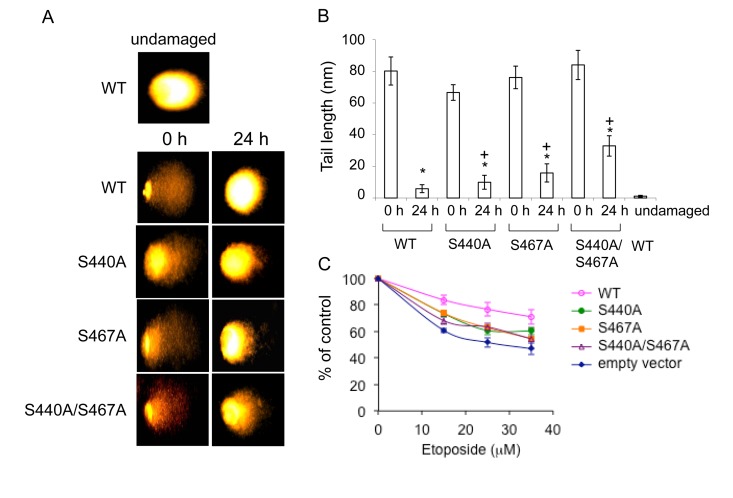
WRN protein, defective in Werner syndrome (WS), a human segmental progeria, is a target of serine/threonine kinases involved in sensing DNA damage. DNA-PK phosphorylates WRN in response to DNA double strand breaks (DSBs). However, the main phosphorylation sites and functional importance of the phosphorylation of WRN has remained unclear. Here, we identify Ser-440 and -467 in WRN as major phosphorylation sites mediated by DNA-PK.In vitro, DNA-PK fails to phosphorylate a GST-WRN fragment with S440A and/or S467A substitution. In addition, full length WRN with the mutation expressed in 293T cells was not phosphorylated in response to DSBs produced by bleomycin. Accumulation of the mutant WRN at the site of laser-induced DSBs occurred with the same kinetics as wild type WRN in live HeLa cells. While the wild type WRN relocalized to the nucleoli after 24 hours recovery from etoposide-induced DSBs, the mutant WRN remained mostly in the nucleoplasm. Consistent with this, WS cells expressing the mutants exhibited less DNA repair efficiency and more sensitivity to etoposide, compared to those expressing wild type. Our findings indicate that phosphorylation of Ser-440 and -467 in WRN are important for relocalization of WRN to nucleoli, and that it is required for efficient DSB repair.


Conflict of interest statement

The authors declare no conflict of interest.

Figures

Figure 1
Figure 1. Mapping DNA-PK phosphorylation sites in WRN
(A) Schematic representation of His- or GST-tagged WRN fragments used in in vitro phosphorylation assay. (B and C) In vitro phosphorylation assay. Purified His- or GST-tagged WRN fragments were incubated with purified DNA-PKcs, Ku 70/86, and activated DNA in the presence of [γ-32P]ATP. Amido black staining is shown (B). The phosphorylation was visualized (C). Asterisk indicates the GST (500-946) fragment. Note that GST (239-499) migrated slower because of many acidic amino acids.
Figure 2
Figure 2. Bleomycin induces WRN phosphorylation at Ser-440 and 467 by DNA-PK
(A-D) In vivo phosphorylation assay with radio-labeling. (A) Empty vector (pEGFP) (lanes 1 and 2) and pEGFP-WRN (lanes 3 and 4) were transfected to 293T cells. The cells were incubated in the absence (lanes 1 and 3) or presence (lanes 2 and 4) of 5 μg/ml bleomycin and [32P] labeled phosphate. WRN proteins were immunoprecipitated. Recombinant His-tagged full length WRN (800 ng) was loaded to assign the position of endogenous WRN (lane 5). The phosphorylated proteins were visualized (upper panel), followed by Western blotting with anti-WRN antibody (lower panel). (B) 293T cells transfected with pEGFP-WRN were treated with 5 μg/ml bleomycin in the presence of PI-3 kinase inhibitors, 25 μM wortmannin (lane 2) and 20 μM NU7026 (lane 3). (C) 293T cells transfected with pEGFP-WRN [wild type (WT) or mutant as indicated] were treated with or without 5 μg/ml bleomycin as indicated. Recombinant His-tagged full length WRN was loaded (lane 7). (D) In vivo phosphorylation assays using anti-phosphoserine antibody. HEK293 cells transfected with pEGFP-WRN (WT or mutant as indicated) were treated with (lanes 5-8) or without 5 μg/ml bleomycin (lanes 1-4),. EGFP proteins were immunoprecipitated with anti-GFP antibody. The phosphorylated proteins were detected by Western blotting with anti-phosphoserine antibody. The membrane was deprobed and analyzed by Western blotting with anti-WRN antibody.
Figure 3
Figure 3. In vitro phosphorylation at Ser-440 and −467 by DNA-PK
GST-tagged WRN fragment (239-499) as schematically represented was used. Purified GST or GST-tagged fragment with or without Ala substitution at Ser-440 and/or −467 was incubated with purified DNA-PKcs, Ku 70/86, and activated DNA in the presence of [γ-32P]ATP. Phosphorylation was visualized (upper panel). An immunoblot with anti-GST antibody is shown (lower panel).
Figure 4
Figure 4. Accumulation of WRN wild type and phosphorylation mutants at laser-induced DSBs
HeLa cells overexpressing either EGFP-WRN wild type (WT) or mutant (S440A, S467A or S440A/S467A) were laser-irradiated at the sites indicated by arrows. Time-dependent accumulation of EGFP-WRN WT and mutants at the DSBs sites were shown.
Figure 5
Figure 5. WRN but not phosphorylation mutant relocalizes to the nucleoli post etoposide exposure
(A) AG11395 cells overexpressing either EGFP-WRN wild type (WT) or mutant (S440A, S467A or S440A/S467A) were incubated with 35 μM etposide for 3 hours. Cells were fixed and EGFP signals were visualized before and after incubation for another 24 hours in fresh medium. Representive images are shown. (B) The percent of cells containing WRN foci. At least 100 cells were scored at each time point. The average of three independent experiments with standard deviation is plotted. Asterisks (*) indicate significant difference between 0 h and 24 h (p<0.05). Plus (+) indicate significant difference between Wild type and mutants.
Figure 6
Figure 6. Cells overexpressing the phosphorylation mutants are moderately sensitive to etoposide
(A) Comet assay with AG11395 cells overexpressing either EGFP-WRN wild type (WT) or mutant (S440A, S467A or S440A/S467A) 24 h after 35 μM etoposide treatment. Representive images are shown. (B) Tail length in (A) are indicated. At least 17 cells were measured. Asterisks (*) indicate significant difference between 0 h and 24 h (p<0.05). Plus (+) indicate significant difference between Wild type and mutants. (C) AG11395 cells overexpressing either EGFP-WRN wild type (WT), S440A, S467A or S440A/S467A were treated with 0, 15, 25 or 35 μM etoposide. 24 h after the treatment, cell proliferation was evaluated by the MTT assay. Significant difference between cells expressing empty vector and WT, or three mutants at concentration of 35 μM (p<0.05).

References

    1. Goto M. Werner's syndrome: from clinics to genetics. Clin Exp Rheumatol. 2000;18:760–766. - PubMed
    1. Martin GM, Sprague CA, Epstein CJ. Replicative life-span of cultivated human cells. Effects of donor's age, tissue, and genotype. Lab Invest. 1970;23:86–92. - PubMed
    1. Salk D, Au K, Hoehn H, Martin GM. Cytogenetics of Werner's syndrome cultured skin fibroblasts: variegated translocation mosaicism. Cytogenet Cell Genet. 1981;30:92–107. - PubMed
    1. Lebel M, Leder P. A deletion within the murine Werner syndrome helicase induces sensitivity to inhibitors of topoisomerase and loss of cellular proliferative capacity. Proc Natl Acad Sci U S A. 1998;95:13097–13102. - PMC - PubMed
    1. Yannone SM, Roy S, Chan DW, Murphy MB, Huang S, Campisi J, et al. Werner syndrome protein is regulated and phosphorylated by DNA-dependent protein kinase. J Biol Chem. 2001;276:38242–38248. - PubMed

Publication types

MeSH terms