. 2014 Mar 7;14(5):972-978.
doi: 10.1039/c3lc50959a.

On-demand, competing gradient arrays for neutrophil chemotaxis

Affiliations

On-demand, competing gradient arrays for neutrophil chemotaxis

Hansang Cho et al. Lab Chip. .

Abstract

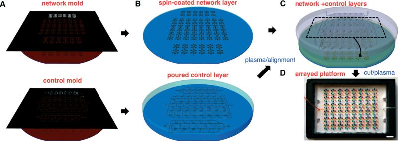
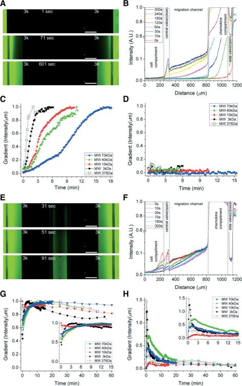
Neutrophils are the most abundant type of white blood cells in the circulation, protecting the body against pathogens and responding early to inflammation. Although we understand how neutrophils respond to individual stimuli, we know less about how they prioritize between competing signals or respond to combinational signals. This situation is due in part to the lack of adequate experimental systems to provide signals in controlled spatial and temporal fashion. To address these limitations, we designed a platform for generating on-demand, competing chemical gradients and for monitoring neutrophil migration. On this platform, we implemented forty-eight assays generating independent gradients and employed synchronized valves to control the timing of these gradients. We observed faster activation of neutrophils in response to fMLP than to LTB4 and unveiled for the first time a potentiating effect for fMLP during migration towards LTB4. Our observations, enabled by the new tools, challenge the current paradigm of inhibitory competition between distinct chemoattractant gradients and suggest that human neutrophils are capable of complex integration of chemical signals in their environment.


Figures

Fig. 1
Fig. 1
Wafer-scale fabrication of arrayed chemotaxis platform. A. Standard photolithographic techniques are used to fabricate SU-8 master molds on 6″ silicon wafers, for the network (upper) and control (lower) layers, respectively. B. PDMS-curing agent mixture is spun-coated and poured to create replicas of the network (upper) and control layers (lower), respectively. C. After curing, plasma-treated PDMS layers are aligned using ethanol as a lubricant and then dried to establish contact between the layers. D. Bonded layers of forty-eight platforms are cut and plasma-treated for bonding to a glass-bottomed plate as a single piece with two control lines for large-scale applications. Scale bar, 1 cm.
Fig. 2
Fig. 2
Schematic representations of on-demand and competing chemotaxis assay. A. Chemokines are loaded in both side compartments (CK1 and CK2) and cells are plated in a middle cellular compartment. These compartments are separated by two side valves (default-closed) and one central valve (default-open), respectively. Sinks between those compartments are designed to balance the difference in priming time of chemokine gradients caused by different diffusivity. Inset shows a photo of the platform visualized by food dyes. B. Two side valves are opened shortly to prime chemokine chambers and migration channels with chemokines and then closed to prevent any disturbing convection flow into the migration channels during chemotaxis assay. C. To plate the cells in the central compartment, the central valve is kept closed, sealing the compartments and avoiding early exposure to chemokines. To expose the cells to chemokine gradients and start the chemotaxis, the central valve is released without any pressure. D. The platform operates in two steps: chemokine priming and chemotaxis. (1–2) With opening of the side valves and closing of the central valve, chemokines fill the chemokine chambers and the migration channels. (3–4) With closing of the side valves and opening of the central valve, chemokines release and activate cell chemotaxis. Scale bars, 2 mm (A) and 200 μm (D).
Fig. 3
Fig. 3
Validation of the initiation and stabilization of gradients for on-demand chemotaxis assay. A–B. Priming the devices and the formation of chemical gradients inside the devices was visualized using FITC-labelled dextran (MW 3 kDa). C. After opening the side valves, chemokine gradients along the migration channels are formed and stabilized within 2 to 15 minutes. Results are presented for fluorescein and various dextran-conjugated dyes, with various molecular weights, ranging from 376 to 70 000 Da. D. The central compartment remained chemokine-free during the priming period. E–F. The initiation of the chemokine gradients was visualized using FITC-labelled dextran (MW 3 kDa). G. After opening the central valve, chemokine gradients along the migration channels were formed and stabilized within 5 minutes. These gradients remained above 80% of the initial slope for over an hour. H. Chemokine gradients along the central compartment also stabilized within 10 minutes and were relatively stable for over an hour. Scale bars, 200 μm.
Fig. 4
Fig. 4
Dominant chemotactic response of human neutrophils to fMLP gradients. A. Individual neutrophils were tracked in fluorescent microscope (left panel: time 0 min, right panel: time 60 min) and their chemotactic activity is compared under dual gradients of fMLP at 100 nM and LTB4 at 100 nM. B. No significant differences in migration speed exist between chemokines. C. Migration toward the fMLP chamber begins earlier than toward the LTB4 chamber and consequently more neutrophils reach the fMLP chambers. D. Neutrophil chemotaxis shows the peak activity in migration speed at 10 nM of fMLP and between 10 to 100 nM of LTB4. E. The neutrophil chemotaxis response time decreases with increasing concentrations of fMLP and LTB4. On average, chemotaxis towards fMLP starts within 15 minutes at 100 nM, ~2 fold faster than towards LTB4 at the same concentration. (Student's t-test. * P < 0.0001 with respect to no-chemokine condition). ncell = 318 for migration speed and ncell = 61 for response time at each concentration. Data represent mean ± s.e.m. Scale bars, 200 μm.
Fig. 5
Fig. 5
Chemotaxis speed and response time in the presence of simultaneous fMLP and LTB4 gradients of various slopes. Contour maps of speed (A) and response time (C) show that chemotaxis towards fMLP is not affected by the presence of LTB4. However, chemotaxis towards LTB4 is affected by the presence of fMLP and the response is faster in the presence of fMLP, both in speed (B) and time (D). ncell = 80 for migration speed and ncell = 15 for response time at each combination of concentration. Data represents mean values (N = 3).

References

    1. Liu M, Chen K, Yoshimura T, Liu Y, Gong W, Wang A, Gao J-L, Murphy PM, Wang JM. Sci. Rep. 2012;2:1–7. - PMC - PubMed
    1. Mackay CR. Nat. Immunol. 2001;2:95–101. - PubMed
    1. Nathan C. Nature. 2002;420:846–852. - PubMed
    1. Arner P. J. Intern. Med. 2007;262:404–407. - PubMed
    1. Serhan CN, Yacoubian S, Yang R. Annu. Rev. Pathol.: Mech. Dis. 2008;3:279–312. - PMC - PubMed

Publication types

MeSH terms

Substances