. 2014 Feb;124(2):670-4.
doi: 10.1172/JCI69519. Epub 2014 Jan 16.

Targeting the cell cycle inhibitor p57Kip2 promotes adult human β cell replication

Targeting the cell cycle inhibitor p57Kip2 promotes adult human β cell replication

Dana Avrahami et al. J Clin Invest. 2014 Feb.

Abstract

Children with focal hyperinsulinism of infancy display a dramatic, non-neoplastic clonal expansion of β cells that have undergone mitotic recombination, resulting in paternal disomy of part of chromosome 11. This disomic region contains imprinted genes, including the gene encoding the cell cycle inhibitor p57Kip2 (CDKN1C), which is silenced as a consequence of the recombination event. We hypothesized that targeting p57Kip2 could stimulate adult human β cell replication. Indeed, when we suppressed CDKN1C expression in human islets obtained from deceased adult organ donors and transplanted them into hyperglycemic, immunodeficient mice, β cell replication increased more than 3-fold. The newly replicated cells retained properties of mature β cells, including the expression of β cell markers such as insulin, PDX1, and NKX6.1. Importantly, these newly replicated cells demonstrated normal glucose-induced calcium influx, further indicating β cell functionality. These findings provide a molecular explanation for the massive β cell replication that occurs in children with focal hyperinsulinism. These data also provided evidence that β cells from older humans, in which baseline replication is negligible, can be coaxed to re-enter and complete the cell cycle while maintaining mature β cell properties. Thus, controlled manipulation of this pathway holds promise for the expansion of β cells in patients with type 2 diabetes.


Figures

Figure 1
Figure 1. Suppression of p57Kip2 in human islets.
(A) Molecular pathology of focal hyperinsulinism. Left: Maternal and paternal copies of chromosome 11 inherited by the fetus. The paternal allele carries a recessive mutation in either ABCC8 or KCNJ11, the two subunits of the ATP-sensitive potassium channel. During fetal life, a break in the maternal chromosome followed by DNA repair using the paternal chromosome as a template occurs in a single β cell. Uniparental disomy of the short arm of chromosome 11 results, as shown in the right panel, and expression of the imprinted p57Kip2 (CDKN1C) gene is silenced. (B) Suppression of p57Kip2 using shRNA lentiviral particles. Quantitative RT-PCR (qPCR) showing mRNA levels of p57Kip2 in HEK293 cells transfected with p57Kip2-specific pGIPZ shRNA constructs. Transfected cells were FACS sorted for GFP-positive and GFP-negative fractions. Expression levels were normalized to an NT shRNA control (n = 3). (C) qRT-PCR of CDKN1C mRNA levels in cultured human islets transduced with lentiviral particles containing pGIPZ shRNA (clone p57-c) against p57Kip2. Five days after transduction, islet were dispersed and FACS sorted for GFP-positive (transduced) and GFP-negative fractions. Expression levels were normalized to NT shRNA control (n = 3 human islet donors). (D) Immunostaining of human islets transduced with pGIPZ shRNA lentivirus against p57Kip2. Turbo-GFP (green), insulin (white), and DNA (blue). Arrows point to costaining of insulin and turbo-GFP. (E) Immunostaining of recovered human islet grafts for turbo-GFP, which allows for tracking of shRNA expression. Turbo-GFP (green), PDX1 (white), and DNA (blue). Arrows point to turbo-GFP/PDX1–positive cells. Scale bars: 20 μm.
Figure 2
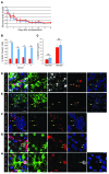
Figure 2. Increased β cell replication in p57Kip2-suppressed human islets.
(A) Blood glucose levels of STZ-treated immunodeficient mice transplanted with p57Kip2-suppressed (blue line) and control human islets (red line) were not statistically different (n = 9 mice per group). (B) BrdU-positive β cells in shRNA lentiviral–transduced, engrafted human islets. Average percentage of BrdU-positive β cells in p57Kip2-suppressed islets (blue bars, 2.71%) was significantly higher (*P < 0.01) than in NT shRNA–transduced islets (red bars, 0.84%). (C) Human and mouse C-peptide levels of mice transplanted with p57Kip2-suppressed and control human islets (blue and red bars, respectively) just before graft recovery (n = 6 per group). (D) Proliferating β cells in p57Kip2-suppressed human islets show negligible p57Kip2 levels. Costaining for p57Kip2 (white), insulin (green), BrdU (red), and DNA (blue). Arrows point to BrdU-positive/p57Kip2-negative β cells. (E) No activation of the DNA damage response by p57Kip2 suppression was observed. Costaining for γH2AX (white), insulin (green), BrdU (red), and DNA (blue). Arrows point to BrdU-positive/γH2AX-negative β cells. (F) Forced cell cycle entry by overexpression of HNF4α induces the DNA damage response. Costaining for γH2AX (white), BrdU (red), and DNA (blue). Arrows point to BrdU/γH2AX double-positive islet cells. (G) 25% ± 9.5% (SD) of newly replicated β cells are doublets. Costaining for PDX1 (green), BrdU (red), and nuclei (blue). Arrows point to BrdU/PDX1 double-positive β cells in close proximity to one another. (H) Some newly replicated β cells stain for the cell cycle marker Ki67. Costaining for Ki67 (white), insulin (green), BrdU (red), and DNA (blue). Arrow points to a BrdU/insulin/Ki67-positive β cell. Scale bars: 20 μm.
Figure 3
Figure 3. Newly replicated β cells retain their functionality as assessed by glucose-stimulated calcium influx assay.
(A) Dispersed human islet cells retrieved from grafts and loaded with the [Ca2+]i sensor Fura-2 were analyzed for [Ca2+]i, followed by costaining for insulin (white), turbo-GFP (green), BrdU (red), and DNA (blue). Blue box surrounds a BrdU-negative β cell and red box surrounds a BrdU-positive β cell. (B) Calcium traces corresponding to the two β cells identified in A, showing the change in [Ca2+]i as a response to an increase in extracellular glucose levels. Each graph represents the [Ca2+]i profile of one cell during the course of the experiment. Original magnification, ×400.

References

    1. Aynsley-Green A. Nesidioblastosis of the pancreas in infancy. Dev Med Child Neurol. 1981;23(3):372–379. - PubMed
    1. Thomas PM, et al. Mutations in the sulfonylurea receptor gene in familial persistent hyperinsulinemic hypoglycemia of infancy. Science. 1995;268(5209):426–429. doi: 10.1126/science.7716548. - DOI - PubMed
    1. Nestorowicz A, et al. A nonsense mutation in the inward rectifier potassium channel gene, Kir6.2, is associated with familial hyperinsulinism. Diabetes. 1997;46(11):1743–1748. doi: 10.2337/diab.46.11.1743. - DOI - PubMed
    1. Goossens A, et al. Diffuse and focal nesidioblastosis. A clinicopathological study of 24 patients with persistent neonatal hyperinsulinemic hypoglycemia. Am J Surg Pathol. 1989;13(9):766–775. doi: 10.1097/00000478-198909000-00006. - DOI - PubMed
    1. Rahier J, Falt K, Muntefering H, Becker K, Gepts W, Falkmer S. The basic structural lesion of persistent neonatal hypoglycaemia with hyperinsulinism: deficiency of pancreatic D cells or hyperactivity of B cells? Diabetologia. 1984;26(4):282–289. - PubMed

Publication types

MeSH terms