. 2014 Mar;25(6):811-27.
doi: 10.1091/mbc.E13-08-0443. Epub 2014 Jan 15.

Critical roles for multiple formins during cardiac myofibril development and repair

Affiliations

Critical roles for multiple formins during cardiac myofibril development and repair

Michelle Rosado et al. Mol Biol Cell. 2014 Mar.

Abstract

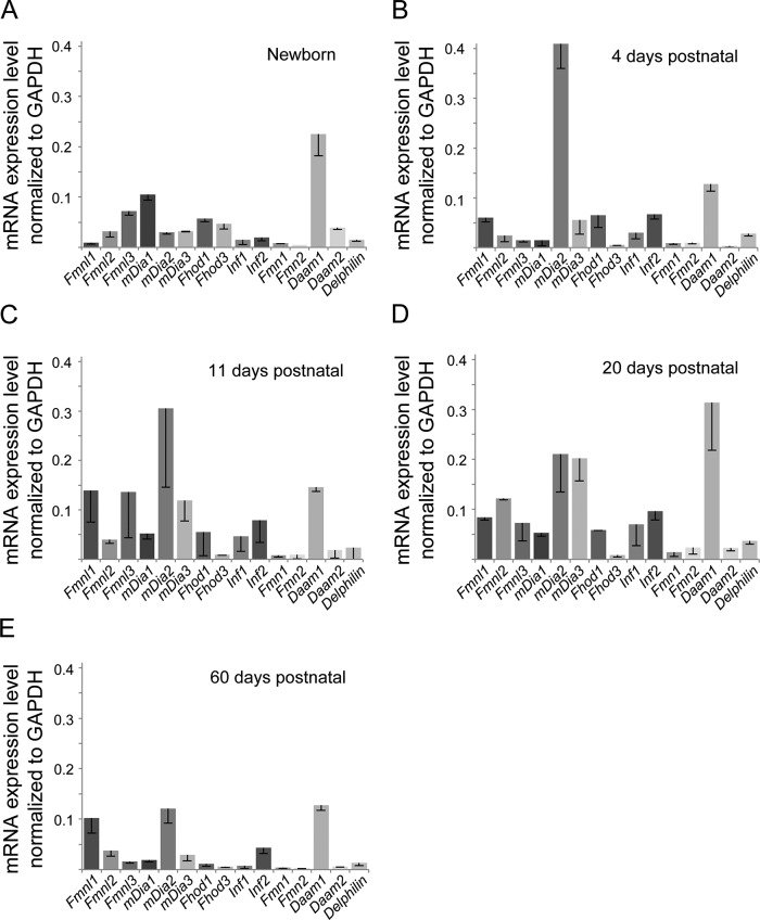
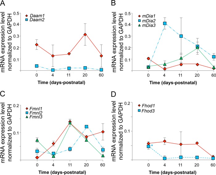
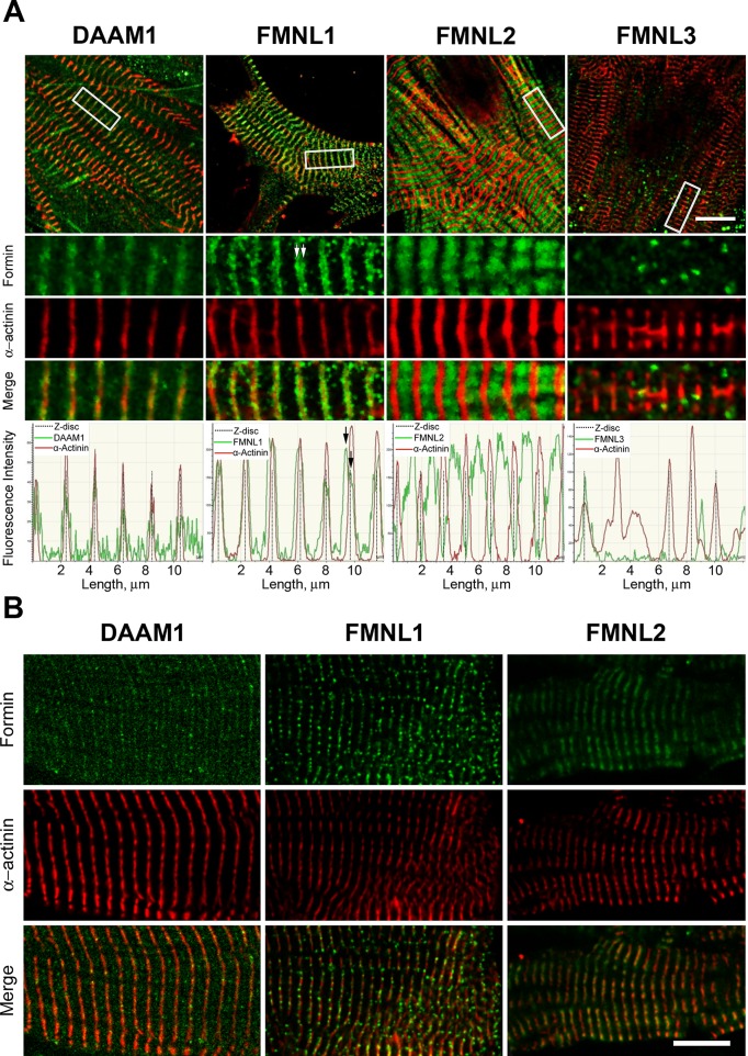
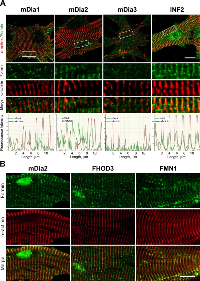
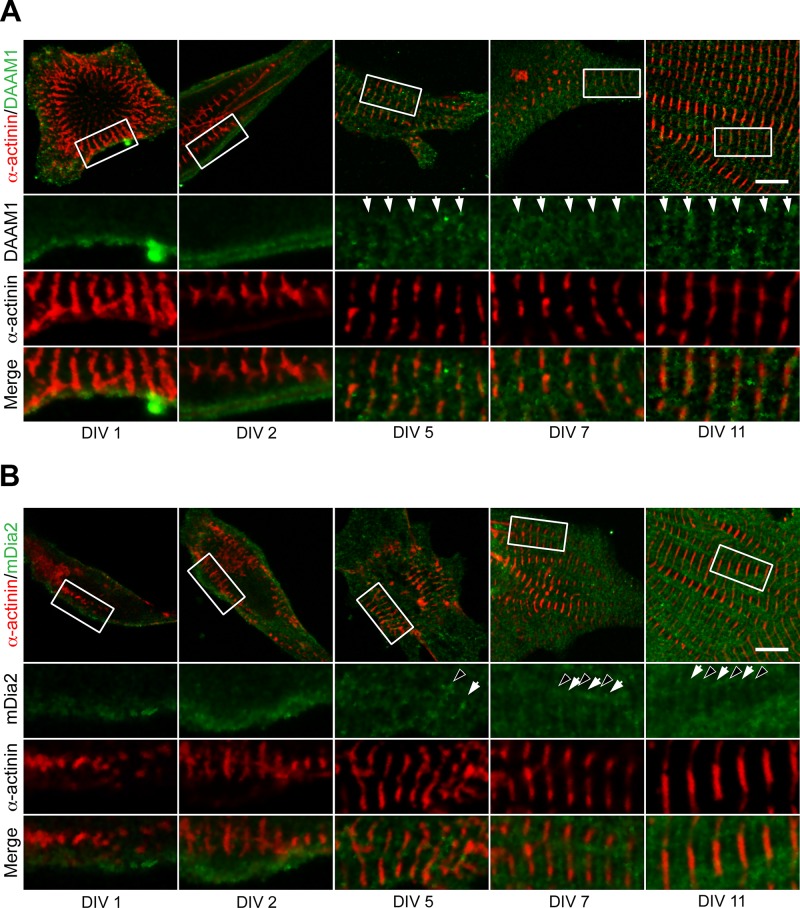
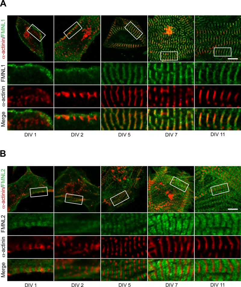
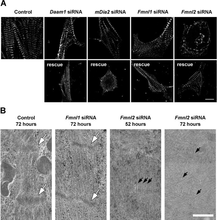
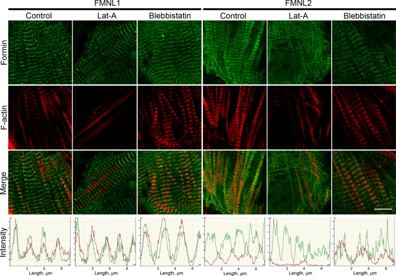
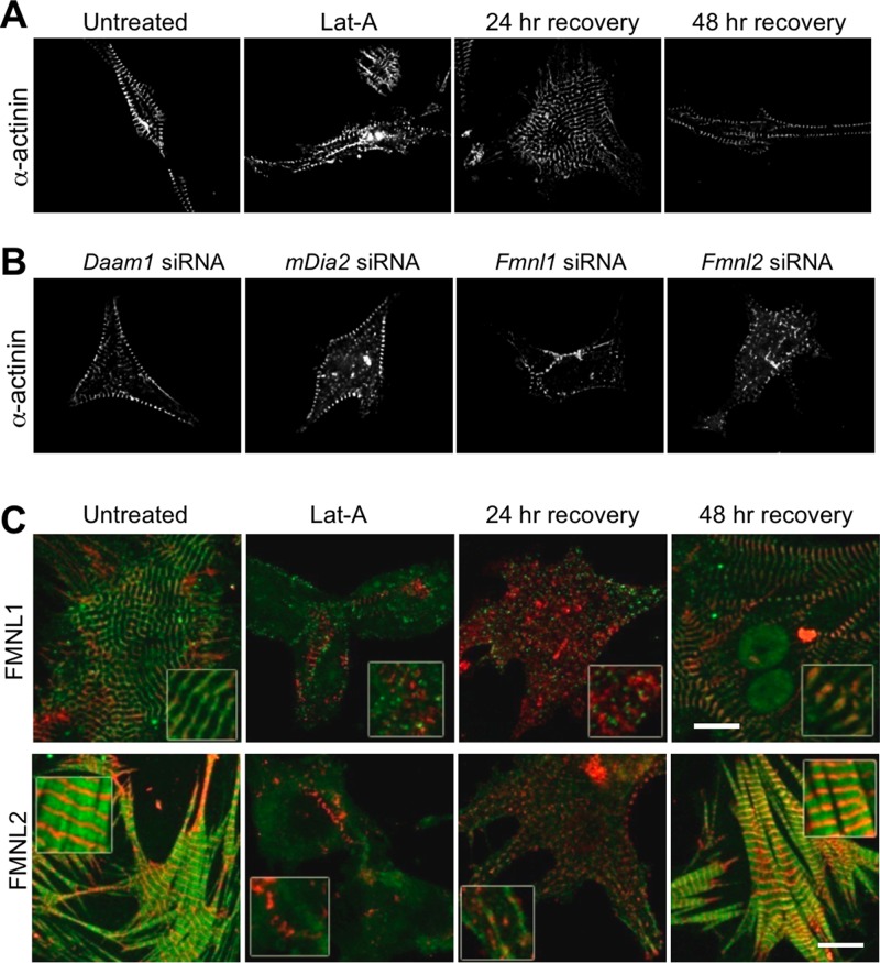
Cardiac and skeletal muscle function depends on the proper formation of myofibrils, which are tandem arrays of highly organized actomyosin contractile units called sarcomeres. How the architecture of these colossal molecular assemblages is established during development and maintained over the lifetime of an animal is poorly understood. We investigate the potential roles in myofibril formation and repair of formin proteins, which are encoded by 15 different genes in mammals. Using quantitative real-time PCR analysis, we find that 13 formins are differentially expressed in mouse hearts during postnatal development. Seven formins immunolocalize to sarcomeres in diverse patterns, suggesting that they have a variety of functional roles. Using RNA interference silencing, we find that the formins mDia2, DAAM1, FMNL1, and FMNL2 are required nonredundantly for myofibrillogenesis. Knockdown phenotypes include global loss of myofibril organization and defective sarcomeric ultrastructure. Finally, our analysis reveals an unanticipated requirement specifically for FMNL1 and FMNL2 in the repair of damaged myofibrils. Together our data reveal an unexpectedly large number of formins, with diverse localization patterns and nonredundant roles, functioning in myofibril development and maintenance, and provide the first evidence of actin assembly factors being required to repair myofibrils.


Figures

FIGURE 1:
FIGURE 1:
Multiple formin genes are differentially expressed during mouse heart development. Comparative qRT-PCR analysis of the mRNA expression levels for the 15 mammalian formins in (A) newborn, (B) 4 d postnatal, (C) 11 d postnatal, (D) 20 d postnatal, and (E) 60 d postnatal mouse hearts. Formin mRNA values were normalized to GAPDH mRNA levels. The formin expression pattern was distinct at each time point. Each bar represents the average value of triplicate reactions from at least three independent experiments. Error bars are SEM.
FIGURE 2:
FIGURE 2:
Developmental changes in formin mRNA levels in mouse heart. Formin mRNA expression levels, as determined by qRT-PCR, throughout postnatal mouse heart development are graphed for members of the (A) DAAM, (B) DIA, (C) FMNL, and (D) FHOD subfamilies. Formin mRNA values were normalized to GAPDH mRNA levels.
FIGURE 3:
FIGURE 3:
Localization of DAAM and FMNL formin subfamily members to sarcomeres. (A) Immunohistochemical localization of the formins (green) DAAM1, FMNL1, FMNL2, and FMNL3 in relation to Z-bands (red, α-actinin) in neonatal primary mouse cardiomyocytes cultured 11 d in vitro. White boxes on cell overview (first row) indicate the zoom-in regions (rows 2–4). Graphs below depict fluorescence intensity in the two channels over 12 μm. Daam1 signal colocalized with α-actinin labeling of the Z-band. FMNL1 signal appeared as a broad band surrounding the α-actinin–labeled Z-band, with peaks on either side of the Z-band (white arrows in zoom; black arrows in graph). FMNL2 signal filled the area between α-actinin–labeled Z-bands. FMNL3 signal appeared as a few puncta with no regular sarcomere-related pattern. (B) Immunohistochemical localization of formin (green) and α-actinin (red) for DAAM1, FMNL1, and FMNL2 in mature myofibrils isolated from 2-mo-old mice. All three formins colocalize with α-actinin, indicating robust sarcomere localization. Scale bars, 10 μm.
FIGURE 4:
FIGURE 4:
Localization of DIA, INF, FHOD, and FMN formin subfamily members to sarcomeres. (A) Immuno­histochemical localization of the formins (green) mDia1, mDia2, mDia3, and INF2 in relation to Z-bands (red, α-actinin) in neonatal primary mouse cardiomyocytes cultured 11 d in vitro. White boxes on cell overview (first row) indicate the zoom-in regions (rows 2–4). Graphs below depict fluorescence intensity in the two channels over 12 μm. mDia1 signal appeared as puncta with no clear sarcomere-related pattern. mDia2 localized to the M-band and Z-band, midway between the α-actinin labeled Z-bands. mDia3 signal was indistinguishable from noise. INF2 colocalized with α-actinin–labeled Z-bands. (B) Immunohistochemical localization of formins (green) and α-actinin (red) for mDia2, FHOD3, and FMN1 in mature myofibrils isolated from 2-mo-old male mice. All three formins colocalize with α-actinin, indicating robust sarcomere localization. Scale bars, 10 μm.
FIGURE 5:
FIGURE 5:
DAAM1 and mDia2 recruitment to sarcomeres during myofibrillogenesis. Immunohistochemistry was used to determine the localization of formins (green) and α-actinin (red) during myofibrillogenesis in cultured neonatal primary cardiomyocytes. Cells were imaged at 1, 2, 5, 7, and 11 DIV. White boxes on cell overview (first row) indicate zoom-in regions (rows 2–4). (A) DAAM1 localized to the cell cortex at DIV 1 and DIV 2, with no apparent sarcomere-related pattern. From DIV 5 onward, DAAM1 appeared in bands (white arrows) that colocalized with α-actinin–labeled Z-bands. (B) mDia2 also localized to the cell cortex at DIV 1 and DIV 2, with no apparent sarcomere-related pattern. At DIV 5, mDia2 began to appear as bands that correlated with the Z-bands (white arrows) or M-bands (black arrowheads). Z-band and M-band staining for mDia2 was robustly observed from DIV 7 onward. Scale bars, 10 μm.
FIGURE 6:
FIGURE 6:
FMNL1 and FMNL2 recruitment to sarcomeres during myofibrillogenesis. Immunohistochemistry was used to determine the localization of formins (green) and α-actinin (red) during myofibrillogenesis in cultured neonatal primary cardiomyocytes. Cells were imaged at 1, 2, 5, 7, and 11 DIV. White boxes on cell overview (first row) indicate zoom-in regions (rows 2–4). (A) At DIV 1–2, FMNL1 localized to the cell cortex near the growing α-actinin–labeled Z-bodies/bands. From DIV 5 onward, FMNL1 localized to broad bands surrounding the Z-bands. (B) FMNL2 localized to clear sarcomere-associated bands between the α-actinin–labeled Z-bands as early as DIV 1. Scale bars, 10 μm.
FIGURE 7:
FIGURE 7:
RNAi silencing of Daam1, mDia2, Fmnl1, or Fmnl2 in cardiomyocytes disrupts myofibril and sarcomere organization. siRNA constructs were used to individually silence Daam1, mDia2, Fmnl1, or Fmnl2 in cultured neonatal primary cardiomyocytes. (A) α-actinin staining of cardiomyocytes 72 h posttransfection revealed structural disruption of myofibrils (top). The absence of the targeted formin protein was confirmed by immunohistochemistry (Supplemental Figure S2). Specificity of the knockdowns was further demonstrated by successful rescues to wild-type phenotype with corresponding siRNA-resistant formin genes (bottom). Scale bar, 10 μm. (B) Electron microscopy imaging of control and siRNA-silenced cells at 52 and 72 h posttransfection. At 72 h posttransfection, siRNA Fmnl1 sarcomeres were longer on average and the Z-bands (white arrows) were thinner on average than in control cells (see Results). By 72 h posttransfection, siRNA Fmnl2 cells contained no structures resembling sarcomeres, although individual disorganized actin filaments were observed (black arrows). At 52 h posttransfection, siRNA Fmnl2 cells contained organized actin filaments but without Z-bands. Scale bar, 500 nm.
FIGURE 8:
FIGURE 8:
FMNL1 and FMNL2 localization in cardiomyocytes after LatA or blebbistatin treatment. After 6 d in culture, neonatal mouse cardiomyocytes were treated with LatA (30 min) or blebbistatin (15 min), fixed, and stained with antibodies. Localization of FMNL1 (green) and F-actin (red) was unaffected by LatA or blebbistatin treatment. Localization of FMNL2 (green) bands was unaffected by LatA treatment. However, blebbistatin caused the broad FMNL2 bands between Z-bands to collapse into narrow bands on either side of the Z-bands. Graphs show fluorescence intensity in the two channels. Scale bars, 10 μm.
FIGURE 9:
FIGURE 9:
FMNL1 and FMNL2 are required for repair of damaged myofibrils. (A) Time course of Z-band repair after injury. To induce myofibril damage, neonatal mouse cardiomyocytes were grown in culture for 48 h, then treated with LatA for 24 h and allowed to recover for 24 or 48 h. α-Actinin banding patterns returned to normal by 24 h of recovery from LatA. (B) Formin requirement in myofibril repair. LatA was used to induce myofibril damage in neonatal primary culture cardiomyocytes in which either Daam1, mDia2, Fmnl1, or Fmnl2 was silenced using siRNA. Images of the injured siRNA cells after 48 h of recovery from LatA treatment. Note that Fmnl1 and Fmnl2 siRNA cells fail to reestablish organized sarcomeric structures, even after 48 h of recovery, whereas Daam1 and mDia2 siRNA cells recovered normally within 24 h. (C) Localization of FMNL1 or FMNL2 (green) and α-actinin (red) in control (untreated) cells, in cells treated with LatA for 24 h, and in cells allowed to recover for 24 or 48 h after LatA treatment. Merged images are shown (individual images are shown in Supplemental Figure S4). Scale bars, 10 μm.

Similar articles

Cited by

References

    1. Arber S, Hunter JJ, Ross J, Hongo M, Sansig G, Borg J, Perriard JC, Chien KR, Caroni P. MLP-deficient mice exhibit a disruption of cardiac cytoarchitectural organization, dilated cardiomyopathy, and heart failure. Cell. 1997;88:393–403. - PubMed
    1. Ayscough KR, Stryker J, Pokala N, Sanders M, Crews P, Drubin DG. High rates of actin filament turnover in budding yeast and roles for actin in establishment and maintenance of cell polarity revealed using the actin inhibitor latrunculin-A. J Cell Biol. 1997;137:399–416. - PMC - PubMed
    1. Breitsprecher D, Goode BL. Formins at a glance. J Cell Sci. 2013;126:1–7. - PMC - PubMed
    1. Campellone KG, Welch MD. A nucleator arms race: cellular control of actin assembly. Nat Rev Mol Cell Biol. 2010;11:237–251. - PMC - PubMed
    1. Chereau D, Boczkowska M, Skwarek-Maruszewska A, Fujiwara I, Hayes DB, Rebowski G, Lappalainen P, Pollard TD, Dominguez R. Leiomodin is an actin filament nucleator in muscle cells. Science. 2008;320:239–243. - PMC - PubMed

Publication types

MeSH terms

LinkOut - more resources