. 2014 Jan 15;6(219):219ra7.
doi: 10.1126/scitranslmed.3007563.

Therapeutic inflammatory monocyte modulation using immune-modifying microparticles

Affiliations

Therapeutic inflammatory monocyte modulation using immune-modifying microparticles

Daniel R Getts et al. Sci Transl Med. .

Abstract

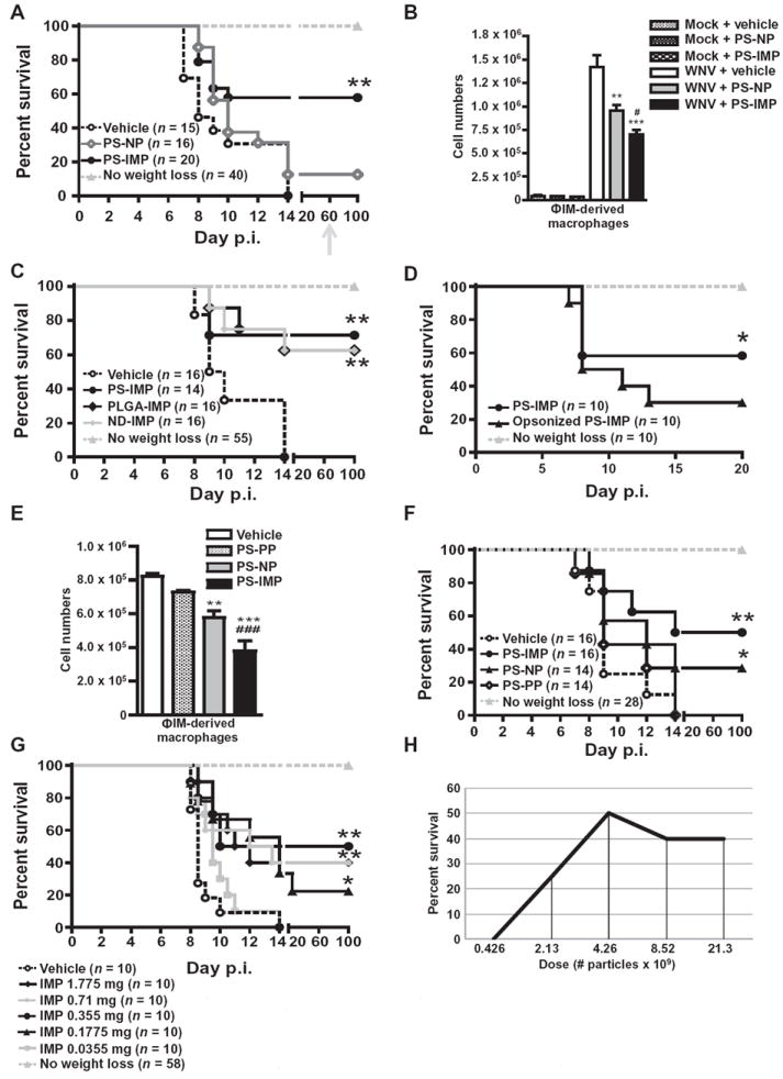
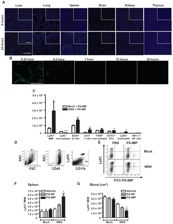
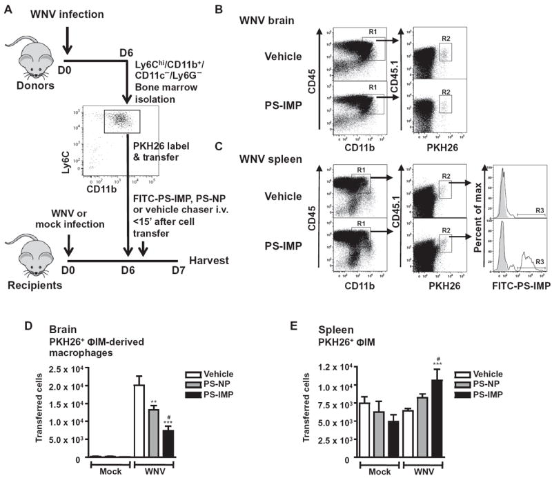
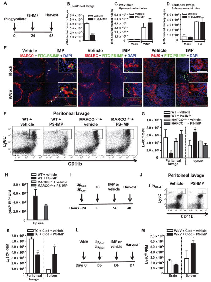
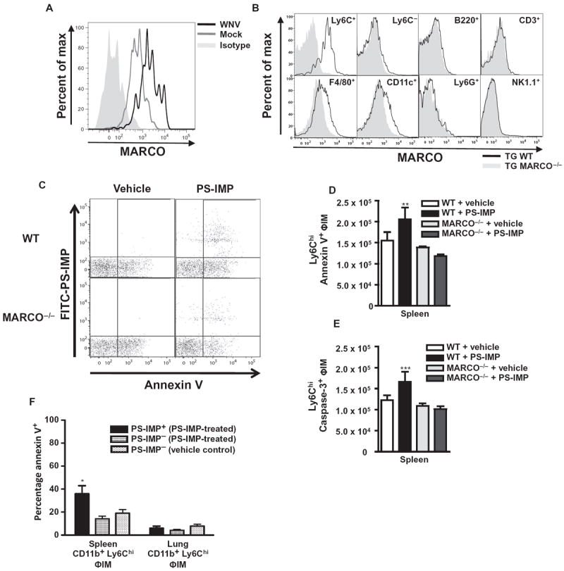
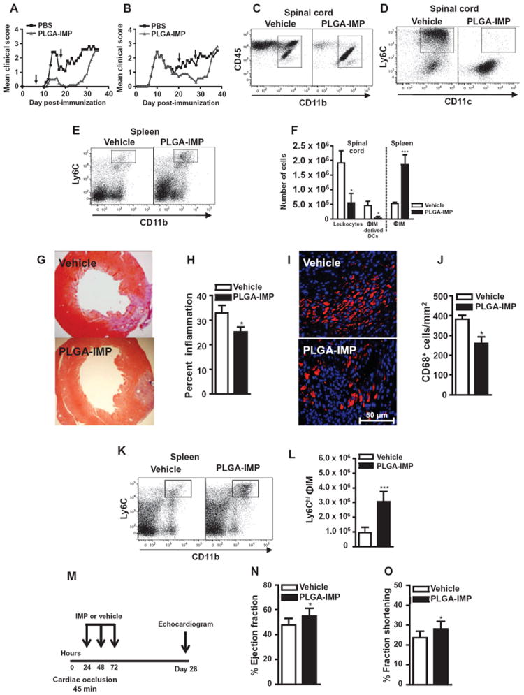
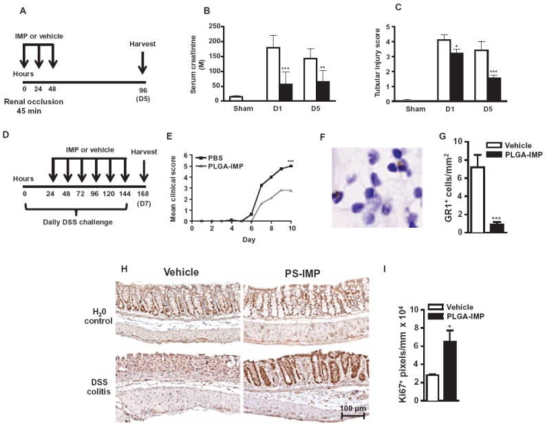
Inflammatory monocyte-derived effector cells play an important role in the pathogenesis of numerous inflammatory diseases. However, no treatment option exists that is capable of modulating these cells specifically. We show that infused negatively charged, immune-modifying microparticles (IMPs), derived from polystyrene, microdiamonds, or biodegradable poly(lactic-co-glycolic) acid, were taken up by inflammatory monocytes, in an opsonin-independent fashion, via the macrophage receptor with collagenous structure (MARCO). Subsequently, these monocytes no longer trafficked to sites of inflammation; rather, IMP infusion caused their sequestration in the spleen through apoptotic cell clearance mechanisms and, ultimately, caspase-3-mediated apoptosis. Administration of IMPs in mouse models of myocardial infarction, experimental autoimmune encephalomyelitis, dextran sodium sulfate-induced colitis, thioglycollate-induced peritonitis, and lethal flavivirus encephalitis markedly reduced monocyte accumulation at inflammatory foci, reduced disease symptoms, and promoted tissue repair. Together, these data highlight the intricate interplay between scavenger receptors, the spleen, and inflammatory monocyte function and support the translation of IMPs for therapeutic use in diseases caused or potentiated by inflammatory monocytes.


Conflict of interest statement

Competing interests: D.R.G., N.J.C.K., and R.L.T. are co-authors on a patent application for modified immune-modulating nanoparticles (#WO2012065153A2). D.R.G., N.J.C.K., S.D.M., and L.D.S. consult for Cour Pharmaceutical Development Company, the license holder of this patent application. These authors are also stakeholders in Cour Pharmaceutical Development Company.

Figures

Fig. 1
Fig. 1. PS-IMPs ameliorate WNV encephalitis in mice
(A) Survival of WNV-infected mice treated with PS-IMPs, PS-NPs (neutral particle control), or vehicle control. Surviving mice were rechallenged with lethal 3 × 10⁸ plaque-forming units (PFU) of WNV on day 60 (gray arrow). (B) Numbers of ΦIM-derived macrophages (SSClo/FSClo/CD45hi/CD11b+/Ly6C+/hi) in the brains of PS-IMP, PS-NP, or vehicle control–treated mock or WNV-infected mice 24 hours after therapy (day 7 after infection). (C) Survival of WNV-infected mice treated with PLGA-IMP, PS-NP, ND-IMP, or vehicle. (D) Survival of WNV-infected mice treated with PS-IMP, opsonized PS-IMP, or vehicle control. (E) Numbers of ΦIM-derived macrophages in the brains of PS-IMP, PS-NP, PS-PP (positively charged particles), or vehicle control–treated or WNV-infected mice 24 hours after therapy (day 7 after infection). (F) Survival of WNV-infected mice treated with PS-IMP, PS-NP, PS-PP, or vehicle control. (G and H) Survival of WNV-infected mice treated with increasing doses of PS-IMP or vehicle control (G) per dose (mg) or (H) as particle number. Survival data represent three separate experiments with 10 to 20 mice per group. Statistical analysis was conducted with the Mantel-Haenszel log-rank test. Flow cytometry data are means ± SD and represent three separate experiments with four to five mice per group. Statistical analysis was conducted with one-way analysis of variance (ANOVA) and Tukey-Kramer post-test. *P ≤ 0.05, **P ≤ 0.01, ***P ≤ 0.001, comparing PS-IMP and NP groups to vehicle control groups; #P ≤ 0.05, ##P ≤ 0.01, ###P ≤ 0.001, comparing PS-IMP to NP groups.
Fig. 2
Fig. 2. PS-IMP treatment redirects inflammatory monocytes to mouse spleen
(A and B) Distribution of FITC–PS-IMPs (green) in the brain, kidney, lung, thymus, spleen, and liver. (B) After intravenous injection of FITC–PLGA-IMPs (green), mouse spleens were processed for fluorescent immunohistology at the time points indicated. (C) Mock- and WNV-infected mice were injected with FITC–PS-IMPs intravenously on day 6 after infection, and spleens were processed for flow cytometry on day 7 after infection to determine localization of PS-IMPs in CD11b+ Ly6C+ inflammatory monocytes (ΦIM), Ly6C macrophages, ΦIM, B220+ B cells, CD3+ T cells, F4/80+ macrophages, CD11c+ DCs, Ly6G+ neutrophils, and NK1.1+ NK cells. (D to G) With flow cytometry, CD45+ Ly6G Ly6Chi monocytes were gated progressively and enumerated in the spleens (F) and blood (G) of mock- and WNV-infected mice 24 hours after FITC–PS-IMP infusion. Immunohistochemistry data represent three separate experiments with three mice per group. Flow cytometry data are means ± SD and represent three separate experiments with four to five mice per group. Statistical analysis was conducted with one-way ANOVA and Tukey-Kramer post-test. *P ≤ 0.05, **P ≤ 0.01, ***P ≤ 0.001, comparing PS-IMP and NP to vehicle control groups; #P ≤ 0.05, ##P ≤ 0.01, ###P ≤ 0.001, comparing PS-IMP to NP groups. (A and B) Scale bars, 200 μm (main photomicrograph) and 50 μm (inset).
Fig. 3
Fig. 3. PS-IMP treatment reduces migration of inflammatory monocytes into the mouse CNS
(A) SSClo/FSClo/CD11b+/CD11c/Ly6Chi inflammatory monocytes (ΦIM) were isolated from the bone marrow of mock- and WNV-infected mouse donors on day 6 after infection, labeled with PKH26 fluorescent dye, and injected into matched recipients on day 6 after infection, immediately followed by a separate injection of vehicle, FITC–PS-NP, or FITC–PS-IMP. (B to E) Brains (B and D) and spleens (C and E) were processed on day 7 after infection for flow cytometry. (B and D) SSClo/FSClo/CD45hi/CD11b+ macrophages (R1) and adoptively transferred PKH26 (R2) populations from brain tissue were progressively gated (B), and transferred cells were enumerated (D). In the spleen (C and E), CD11c/CD45hi/CD11b+ macrophages (R1), adoptively transferred PKH26 (R2), and IMP+ macrophage populations (R3) were progressively gated for examination (B), with transferred cells enumerated in (E). Flow cytometry data are means ± SD and represent three separate experiments with four to five mice per group. Statistical analysis was conducted with one-way ANOVA and Tukey-Kramer post-test. *P ≤ 0.05, **P ≤ 0.01, ***P ≤ 0.001, comparing PS-IMP and NP to vehicle control groups; #P ≤ 0.05, ##P ≤ 0.01, ###P ≤ 0.001, comparing PS-IMP and NP groups.
Fig. 4
Fig. 4. The scavenger receptor MARCO mediates IMP activity
(A and B) PLGA-IMP or vehicle (A) was injected 24 hours after TG injection, with the number of peritoneal CD11b+/Ly6Chi/Ly6G inflammatory monocytes (ΦIM) enumerated by flow cytometry (B). (C and D) The importance of the spleen for IMP efficacy was addressed in splenectomized mice, with ΦIM numbers in the brains of WNV-infected mice (C) or the peritoneal cavity (D) of mice with TG-induced peritonitis compared between vehicle and IMP treatment. (E) FITC–PS-IMPs (green) or vehicle control were infused on day 6 after WNV or mock infection and processed 24 hours later for expression of MARCO, SIGLEC, or F4/80 immunohistology with 4′,6-diamidino-2-phenylindole (DAPI) (blue) counterstaining. White arrowheads indicate co-localization between MARCO+ cells and FITC–PS-IMP. (F and G) PS-IMP or vehicle was injected intravenously into wild-type (WT) or MARCO−/− mice 24 hours after TG injection, with total peritoneal inflammation examined by flow cytometry (F) and the total number of ΦIM-derived macrophages determined (G). (H) In addition, both total (G) and PS-IMP+ (H) Ly6Chi ΦIM macrophages were enumerated in the spleen. (I to M) Deletion of marginal zone macrophages was performed with clodronate liposomes, and IMP efficacy in mice with TG-induced peritonitis (I to K) or WNV encephalitis (L and M) was examined. In TG-treated animals (I), ΦIM-derived macrophages in the peritoneal cavity and spleen were enumerated (J and K) by flow cytometry. In WNV-infected mice (L), Ly6Chi ΦIM-derived macrophages in the brain and spleen were examined by flow cytometry on day 7 after infection (M). Flow cytometry data are means ± SD and represent three separate experiments with four to five mice per group. Statistical analysis was conducted with one-way ANOVA and Tukey-Kramer post-test. *P ≤ 0.05, **P ≤ 0.01, ***P ≤ 0.001, comparing the WT PS-IMP groups to all other groups. Immunohistochemistry data represent three separate experiments with three mice per group. (E) Scale bars, 200 μm (main photomicrograph) and 50 μm (inset).
Fig. 5
Fig. 5. Uptake of PS-IMPs by MARCO+ inflammatory monocytes induces apoptosis
(A and B) MARCO expression on peripheral blood inflammatory monocytes (ΦIM) from WNV-infected animals at day 7 after infection (A), as well as on peripheral blood leukocyte populations in WT or MARCO−/− mice 24 hours after TG injection (B). (C to E) With the TG model of colitis, the numbers of splenic annexin V+ CD11b+ Ly6Chi Ly6G ΦIM (C and D) and caspase-3+ CD11b+ Ly6Chi Ly6G ΦIM (E) were examined. (F) In the spleen and lung, the percentage of annexin V+ CD11b+ Ly6Chi Ly6G ΦIM that were PS-IMP+ or PS-IMP was compared. Flow cytometry data are means ± SD and represent three separate experiments with four to five mice per group. Statistical analysis was conducted with one-way ANOVA and Tukey-Kramer post-test. *P ≤ 0.05, **P ≤ 0.01, ***P ≤ 0.001, comparing the WT PS-IMP groups to all other groups.
Fig. 6
Fig. 6. PLGA-IMPs ameliorate disease in EAE and cardiac infarction
(A and B) EAE disease scores in mice treated for 10 days with PLGA-IMPs or vehicle at onset (A) or relapse (B). (C to E) CD45+/CD11b+/Ly6Chi/CD11c+ ΦIM-derived DCs in spinal cords (C and D) and CD45+/CD11b+/Ly6Chi ΦIM in spleens (E) of mice treated for 7 days at disease onset. (F) Cell numbers in spinal cord and spleen. (G and H) Cardiac histology (G) and infarction size quantification (H) were performed 7 days after permanent left anterior descending artery occlusion in mice treated with three daily infusions of PLGA-IMPs or vehicle beginning 24 hours after occlusion. (I and J) CD68+ macrophages were enumerated by immunohistochemistry. (K and L) Spleens were processed for flow cytometry 7 days after occlusion, and CD45+/CD11b+/Ly6C+/ΦIM were gated and enumerated. (M to O) Function after temporary cardiac arterial occlusion was determined by echocardiography 28 days after occlusion (M), the percentage cardiac ejection fraction (N), and fraction shortening (O) 28 days after reperfusion. EAE mean clinical score data are representative of three separate experiments with 10 to 20 mice per group. Flow cytometry data are means ± SD and represent three separate experiments with four to five mice per group. Cardiac infarction data are representative of two experiments with at least three mice per group. Image analysis was performed as described in Materials and Methods. Statistical analysis was conducted with unpaired, two-tailed Student’s t test. *P ≤ 0.05, **P ≤ 0.01, ***P ≤ 0.001, comparing PLGA-IMP and vehicle control group.
Fig. 7
Fig. 7. PS-IMP ameliorates disease in kidney injury and colitis
(A to C) PLGA-IMP treatment was tested after temporary renal artery occlusion (A) using serum creatinine (B) and tubular injury score (C) as functional measures at days 1 and 5 after reperfusion. (D) PS-IMP or vehicle control was also injected daily intravenously into mice with DSS-induced colitis or control (water vehicle) from day 1 to day 6 after disease induction. (E to G) Daily mean clinical scores (E) and GR1+ monocytes were enumerated at day 7 (F and G). (H) Mouse colon tissue was processed for immunohistology on day 9 of DSS challenge, and Ki67+ staining (brown) was performed on transverse colon sections. (I) Image analysis was conducted comparing DSS-induced colitis vehicle to PS-IMP. Renal artery occlusion and DSS-induced colitis data are representative of at least three experiments with at least 5 to 20 mice per group. Image analysis was performed as described in Materials and Methods. Statistical analysis was conducted with unpaired, two-tailed Student’s t test. *P ≤ 0.05, **P ≤ 0.01, ***P ≤ 0.001, comparing PLGA-IMP and vehicle control group.

Comment in

Similar articles

Cited by

References

    1. Bain CC, Scott CL, Uronen-Hansson H, Gudjonsson S, Jansson O, Grip O, Guilliams M, Malissen B, Agace WW, Mowat AM. Resident and pro-inflammatory macrophages in the colon represent alternative context-dependent fates of the same Ly6Chi monocyte precursors. Mucosal Immunol. 2013;6:498–510. - PMC - PubMed
    1. Getts DR, Terry RL, Getts MT, Müller M, Rana S, Shrestha B, Radford J, Van Rooijen N, Campbell IL, King NJ. Ly6c+ “inflammatory monocytes” are microglial precursors recruited in a pathogenic manner in West Nile virus encephalitis. J Exp Med. 2008;205:2319–2337. - PMC - PubMed
    1. King IL, Dickendesher TL, Segal BM. Circulating Ly-6C+ myeloid precursors migrate to the CNS and play a pathogenic role during autoimmune demyelinating disease. Blood. 2009;113:3190–3197. - PMC - PubMed
    1. Lin KL, Sweeney S, Kang BD, Ramsburg E, Gunn MD. CCR2-antagonist prophylaxis reduces pulmonary immune pathology and markedly improves survival during influenza infection. J Immunol. 2011;186:508–515. - PMC - PubMed
    1. Schiopu A, Nadig SN, Cotoi OS, Hester J, van Rooijen N, Wood KJ. Inflammatory Ly-6Chi monocytes play an important role in the development of severe transplant arteriosclerosis in hyperlipidemic recipients. Atherosclerosis. 2012;223:291–298. - PMC - PubMed

Publication types

MeSH terms