. 2014 Jan 15;6(219):219ra8.
doi: 10.1126/scitranslmed.3007828.

Human TH9 cells are skin-tropic and have autocrine and paracrine proinflammatory capacity

Affiliations

Human TH9 cells are skin-tropic and have autocrine and paracrine proinflammatory capacity

Christoph Schlapbach et al. Sci Transl Med. .

Abstract

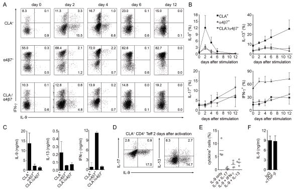
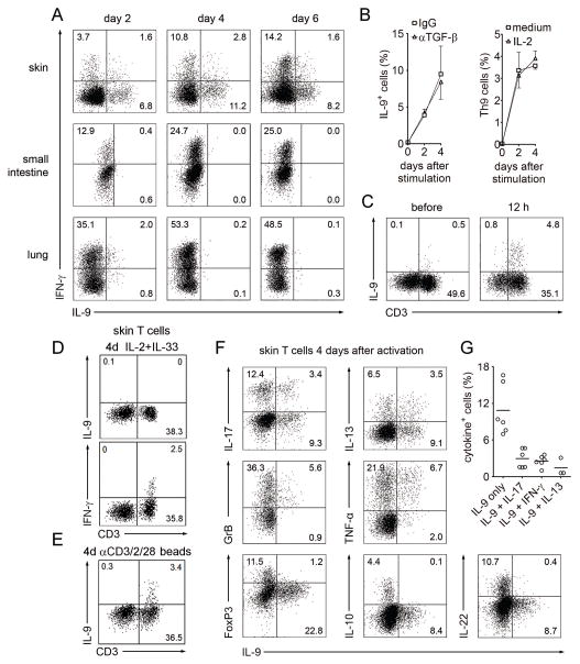
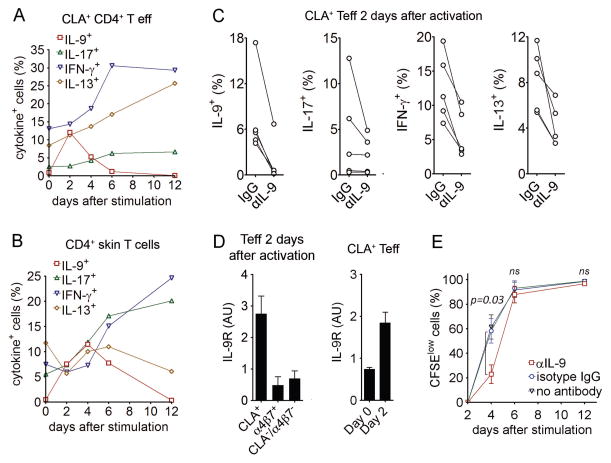
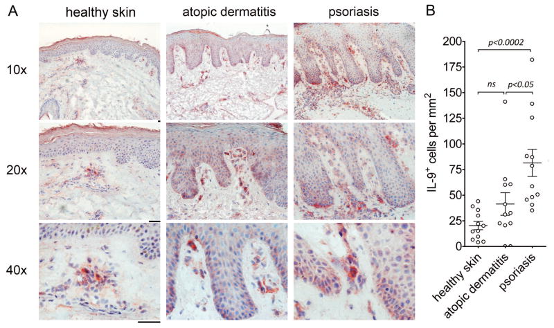
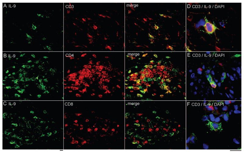
T helper type 9 (TH9) cells can mediate tumor immunity and participate in autoimmune and allergic inflammation in mice, but little is known about the TH9 cells that develop in vivo in humans. We isolated T cells from human blood and tissues and found that most memory TH9 cells were skin-tropic or skin-resident. Human TH9 cells coexpressed tumor necrosis factor-α and granzyme B and lacked coproduction of TH1/TH2/TH17 cytokines, and many were specific for Candida albicans. Interleukin-9 (IL-9) production was transient and preceded the up-regulation of other inflammatory cytokines. Blocking studies demonstrated that IL-9 was required for maximal production of interferon-γ, IL-9, IL-13, and IL-17 by skin-tropic T cells. IL-9-producing T cells were increased in the skin lesions of psoriasis, suggesting that these cells may contribute to human inflammatory skin disease. Our results indicate that human TH9 cells are a discrete T cell subset, many are tropic for the skin, and although they may function normally to protect against extracellular pathogens, aberrant activation of these cells may contribute to inflammatory diseases of the skin.


Conflict of interest statement

CONFLICT OF INTEREST

RAC and TSK previously had an equity interest in TremRX, a start-up company that seeks as a long-term business plan to improve vaccine formulation and delivery. During the period RAC and TSK held the equity, the interest was deemed to create a financial conflict of interest (as defined by the specific Public Health Serivce regulations) with the research discussed in this article. To resolve this matter, RAC and TSK divested themselves of the equity interest in this company, so this financial conflict of interest no longer exists. RAC has served as a consultant for Novartis, Dermira and Stiefel. The other authors declare that they have no competing interests.

Figures

Fig. 1
Fig. 1. IL-9 is transiently and selectively produced by skin-tropic TH cells after stimulation with Candida albicans.
(A) Tissue tropic T cells preferentially respond to pathogens encountered through that epithelial surface. The % maximal proliferation (% max prolif) of T cells tropic for skin (CLA+), gut (α4β7+) and other tissues (CLAα4β7) is shown. CLA+, α4β7+, and CLA/α4β7 memory CD4+ T cells were isolated from PBMC of healthy donors and separately stimulated with autologous monocytes pulsed with a panel of viral, bacterial and fungal antigen preparations. Proliferation was assayed by CFSE dilution and maximal proliferation for each antigen was used to calculate relative responses of other T cell subsets (mean + s.e.m. of duplicates). *p=0.0001, otherwise p values are as shown. (B–E) IL-9 was produced selectively by skin tropic T cells in response to C. albicans. Cytokine production of T cell subsets following stimulation with autologous antigen-pulsed monocytes was measured in cell culture supernatants using a bead-based multiplex assay (mean + s.e.m. of duplicates). Significant IL-9 production was observed only in CLA+ skin tropic T cells stimulated with C. albicans whereas IFN-γ, IL-13 and IL-17 were produced in variable amounts by all tissue tropic subsets and in response to all antigens tested. (F) All tissue tropic T cell subsets proliferated in response to C. albicans, demonstrating that selective IL-9 production by skin tropic T cells was not the result of poor recognition of C. albicans by other T cell subsets. The % CFSElow T cells is shown (mean + s.e.m. of duplicates). (G,H) IL-9 is transiently produced after pathogen stimulation. T cells were stimulated with C. albicans and S. aureus, and IL-9 and IL-17 production was assessed at different time points by flow cytometry after stimulation with phorbol 12-myristate 13-acetate and ionomycin (PMA+I). Cytokine production of stimulated T helper cells on day 6 (G) and the kinetics of IL-9 and IL-17 production (H) are shown (mean + s.d.). Data are representative of independent experiments with 5 (A,F), 3 (B–E) or 2 (G,H) donors.
Fig. 2
Fig. 2. Human TH9 cells are a distinct T cell population tropic for the skin
(A, B) CLA+, α4β7+, and CLA/α4β7 CD25 memory CD4+ T cells (TEFF) were isolated from healthy donors and polyclonally stimulated with αCD3/αCD2/αCD28. The production of IL-9, IFN-γ, IL-13, and IL-17 was assessed by flow cytometry after stimulation with PMA+I at the indicated time points. Histograms from an individual donor (A) and aggregate data are shown (B) (mean + s.d.). The majority of IL-9 production was observed in CLA+ skin tropic T cells and IL-9 was produced only transiently after activation, in contrast to other cytokines tested simultaneously (IL-13, IL-17, IFN-γ). (C) Cytokine production as measured by bead multiplex analysis of supernatants from tissue tropic subsets on day 4 after stimulation is shown (mean + s.d.); results confirmed flow cytometry studies demonstrating most IL-9 is produced by CLA+ T cells. (D,E) Most IL-9 producing T cells lacked production of other TH lineage cytokines. 2 days after stimulation, CLA+ Teff were analyzed by flow cytometry for co-production of IL-9 and IL-17, IFN-γ and IL-13. Representative histograms (D) and aggregate data (E) are shown (mean + s.d.). (F) IL-9 production was not dependent on the presence of TGF-β. IL-9 in supernatants from T cells stimulated for four days in the presence of neutralizing antibody to TGF-β or an isotype-matched control antibody is shown (mean + SD). Data are representative of independent experiments with at least 4 donors.
Fig. 3
Fig. 3. TH9 cells are selectively found in human skin, constitute a distinct T cell population and are independent of TGF-β and IL-2
(A) TH9 cells are resident in human skin but not small intestine or lung. T cells were isolated from healthy human skin, small intestine and lung, stimulated with αCD3/αCD2/αCD28 and production of IL-9 and IFN-γ was assessed by flow cytometry at the indicated time points after stimulation with PMA+I. (B) Skin TH9 cells are independent of TGF-β and IL-2. T cells freshly isolated from healthy skin were stimulated as in (A) and cultured in the presence or absence of neutralizing antibody to TGF-β or isotype matched control antibody (left) or exogenous IL-2 (right). Percentage of IL-9+ Th cells was assessed by flow cytometry at the indicated time points after stimulation with PMA+I (mean + s.d.). (C) CD3+ T cells are the major source of IL-9 in human skin. Cell suspensions from human skin were simulated with PMA+I before or after 12 hours of stimulation with αCD3/αCD2/αCD28 beads. CD3+ T cells were the major source of IL-9 both before and after T cell stimulation. (D,E) Innate lymphoid cells (ILC) were not an appreciable source of IL-9 in healthy human skin. Cell suspensions from human skin were stimulated for four days (4d) with either (D) IL-2 (10 U/ml)+IL-33 (50 ng/ml) (to stimulate ILC) or (E) αCD3/αCD2/αCD28 beads (to stimulate T cells), then treated with PMA+I. IL-9 was not produced by CD3 cells after IL-2+IL-33 but was produced by CD3+ cells after bead treatment. CD3+ T cells produced IFN-γ without bead stimulation but IL-9 production was not observed unless T cells were first bead stimulated. (F) TH9 cells co-produced granzyme B and TNF-α but lacked FoxP3 and production of other TH lineage cytokines. Four days after stimulation, skin resident T cells were analyzed for co-expression of IL-9 with various cytokines, granzyme B and FoxP3. (G) The frequency of skin resident T cells producing IL-9 alone or in combination with IL-17, IFN-γ, or IL-13, 4 days after stimulation is shown. Data are representative of independent experiments with at least 3 (A, D, E) or 6 donors (B, C, F, G). Panels C–E are gated to show all viable cells as determined by forward/side scatter, panels A and F show viable CD3+/CD8 lymphocytes.
Fig. 4
Fig. 4. Human TH9 cells have autocrine and paracrine pro-inflammatory activity
(A,B) IL-9 production is transient and precedes the up-regulation of other inflammatory cytokines. T cells isolated from blood (A) and skin (B) were stimulated with αCD3/αCD2/αCD28 and production of IL-9, IFN-γ, IL-17, and IL-13 was assessed at the indicated time points by flow cytometry after stimulation with PMA+I. (C) IL-9 production is required for maximal production of IL-9 itself as well for as the production of other inflammatory cytokines. CLA+ TEFF were stimulated for 2 days with αCD3/αCD2/αCD28 in the presence of neutralizing antibody to IL-9 or isotype matched control antibody. Cytokine production was assessed by flow cytometry after stimulation with PMA+I. (D) Expression of the IL-9 receptor is enriched in activated skin-tropic CLA+ TEFF. IL-9 receptor (IL-9R) mRNA was measured by real-time quantitative PCR in CLA+, α4β7+, and CLA/α4β7 TEFF after 2 days of stimulation with αCD3/αCD2/αCD28 (mean + s.e.m., 3 donors with triplicates). Baseline expression (Day 0) vs. expression after activation (Day 2) of IL-9R by CLA+ TEFF cells is shown in the right panel. (E) IL-9 enhances cellular proliferation at early timepoints. TEFF were labeled with CFSE, stimulated with αCD3/2/28 with anti-IL-9 (αIL-9) or isotype matched control antibody and proliferative cells were identified (CFSElow). Proliferation was significantly decreased at day four but was unchanged at later time points. The mean and SEM of four donors are shown. Data are representative of independent experiments with 6 (A,B), 5 (C, E), 3 (D) donors.
Fig. 5
Fig. 5. IL-9 producing cells are increased in the skin lesions of psoriasis and atopic dermatitis
(A) Healthy skin and lesional skin samples from patients with psoriasis and atopic dermatitis were immunohistochemically stained for IL-9. Positive cells appear red. (B) TH9 cells are significantly increased in the skin lesions of psoriasis but not atopic dermatitis. The number of IL-9+ cells per mm2 in immunohistochemically stained sections of healthy, atopic dermatitis and psoriasis skin are shown. The mean and SEM of 2 independent experiments with 12 donors each per condition are shown. Scale bars=50 μM.
Fig. 6
Fig. 6. TH9 cells are evident in human psoriatic lesional skin
(A–C) Two color immunofluorescence staining was performed on lesional psoriatic skin. A population of IL-9 producing cells expressed (A) CD3, (B) CD4, and (C) lacked expression of CD8. (D, E) IL-9 production was observed in cells undergoing cell division. (F) A population of CD3 IL-9 producing cells was also present in psoriatic skin lesions. Scale bars=10 μM.

Similar articles

Cited by

References

    1. Dardalhon V, Awasthi A, Kwon H, Galileos G, Gao W, Sobel RA, Mitsdoerffer M, Strom TB, Elyaman W, Ho IC, Khoury S, Oukka M, Kuchroo VK. IL-4 inhibits TGF-beta-induced Foxp3+ T cells and, together with TGF-beta, generates IL-9+ IL-10+ Foxp3(−) effector T cells. Nature immunology. 2008;9:1347–1355. - PMC - PubMed
    1. Veldhoen M, Uyttenhove C, van Snick J, Helmby H, Westendorf A, Buer J, Martin B, Wilhelm C, Stockinger B. Transforming growth factor-beta ‘reprograms’ the differentiation of T helper 2 cells and promotes an interleukin 9-producing subset. Nature immunology. 2008;9:1341–1346. - PubMed
    1. Wilhelm C, Turner JE, Van Snick J, Stockinger B. The many lives of IL-9: a question of survival? Nature immunology. 2012;13:637–641. - PubMed
    1. Purwar R, Schlapbach C, Xiao S, Kang HS, Elyaman W, Jiang X, Jetten AM, Khoury SJ, Fuhlbrigge RC, Kuchroo VK, Clark RA, Kupper TS. Robust tumor immunity to melanoma mediated by interleukin-9-producing T cells. Nature medicine. 2012 - PMC - PubMed
    1. Lu Y, Hong S, Li H, Park J, Hong B, Wang L, Zheng Y, Liu Z, Xu J, He J, Yang J, Qian J, Yi Q. Th9 cells promote antitumor immune responses in vivo. The Journal of clinical investigation. 2012 - PMC - PubMed

Publication types

MeSH terms