Review
. 2014 Feb;6(1):13-26.
doi: 10.1093/jmcb/mjt053. Epub 2014 Jan 14.

Targeting Wnt signaling at the neuroimmune interface for dopaminergic neuroprotection/repair in Parkinson's disease

Affiliations
Review

Targeting Wnt signaling at the neuroimmune interface for dopaminergic neuroprotection/repair in Parkinson's disease

Francesca L'Episcopo et al. J Mol Cell Biol. 2014 Feb.

Abstract

During the past three decades, the Wingless-type MMTV integration site (Wnt) signaling cascade has emerged as an essential system regulating multiple processes in developing and adult brain. Accumulating evidence points to a dysregulation of Wnt signaling in major neurodegenerative pathologies including Parkinson's disease (PD), a common neurodegenerative disorder characterized by the progressive loss of midbrain dopaminergic (mDA) neurons and deregulated activation of astrocytes and microglia. This review highlights the emerging link between Wnt signaling and key inflammatory pathways during mDA neuron damage/repair in PD progression. In particular, we summarize recent evidence documenting that aging and neurotoxicant exposure strongly antagonize Wnt/β-catenin signaling in mDA neurons and subventricular zone (SVZ) neuroprogenitors via astrocyte-microglial interactions. Dysregulation of the crosstalk between Wnt/β-catenin signaling and anti-oxidant/anti-inflammatory pathways delineate novel mechanisms driving the decline of SVZ plasticity with age and the limited nigrostriatal dopaminergic self-repair in PD. These findings hold a promise in developing therapies that target Wnt/β-catenin signaling to enhance endogenous restoration and neuronal outcome in age-dependent diseases, such as PD.

Keywords: Parkinson's disease; Wnt/β-catenin signaling; dopaminergic neurons; neurodegeneration; neurogenesis; neuroinflammation; neuroprotection.


Figures

Figure 1
Figure 1. Wnt signaling cascades.
(A) In canonical Wnt/β-catenin pathway, binding of Wnts (‘Wnt on’) to a receptor complex composed of Fzd and LRP family members inhibits the APC/GSK-3β destruction complex and blockade of β-catenin (β-cat) by GSK-3β. β-cat then accumulates in the cytoplasm and translocates to the nucleus where it regulates target gene expression with TCF/LEF family of transcription factors. In the absence of Wnt ligand (‘Wnt off’), β-cat is targeted for proteolytic degradation by the APC/GSK-3β destruction complex. The proteins Norrin and R-spondin (Rspo) are unrelated to Wnt and act as Wnt agonists, whereas Dkk1, Wif, and sFRPs act as antagonists. (B) In noncanonical Wnt/Ca2+ pathway, the binding of Wnts promotes Fzd-mediated activation of G proteins, leading to the release of Ca2+ from intracellular stores and consequent activation of Ca2+-dependent effector molecules. Several Ca2+-sensitive targets, i.e. PKC, CamKII, and calcineurin, have been identified downstream of the Wnt/Ca2+ pathway. Targets of the Wnt/Ca2+ pathway appear to interact with the Wnt/β-catenin pathway at multiple points. Additionally, Fzd receptors in association with Kny, Ror2, or Ryk receptors can activate JNK, promoting target gene expression through AP-1. In noncanonical Wnt/PCP pathway, the binding of Wnts activates RhoA/B, Cdc42, or Rac1. Dvl activates Rac1, which can activate JNK to signal through the NFAT pathway.
Figure 2
Figure 2. Schematic illustration of Wnt1/β-catenin signaling as a key player in mDA neuron survival/protection.
In the intact midbrain, canonical Wnt1-like agonists via activation of Fzd-1 receptors (‘Wnt on’) maintain the integrity of mDA neurons by blocking GSK-3β-induced phosphorylation (P) and proteosomal degradation of β-catenin. Stabilized β-catenin can translocate into the nucleus and associate with transcription factors to regulate the expression of Wnt target genes involved in DA neuron survival/plasticity. β-catenin may also function as a pivotal defense molecule against oxidative stress or as a coactivator for several nuclear receptors involved in the maintenance/protection of DA neurons (L’Episcopo et al., 2011b). Neurotoxic agents including PD neurotoxins (MPTP/MPP+, 6-OHDA), pesticides (rotenone), increased oxidative load as a result of growth factors (GFs) deprivation, or aging may antagonize Wnt/β-catenin signaling (‘Wnt off’) in DA neurons. Upregulation of active GSK-3β leads to β-catenin degradation and increased DA neuron vulnerability/degeneration/apoptosis. Various potential endogenous Wnt agonists (Respondin, Rspo, Norrin) or antagonists (Dkk1, Wif, sFRP) are also indicated.
Figure 3
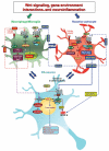
Figure 3. Gene-environment interactions and crosstalk among astrocytes, microglia, and DA neuronsvia Wnt signaling.
Upon nigrostriatal injury, at the neuron–astrocyte interface, astrocyte-derived Wnt1 via Fzd-1 receptor likely transmits pro-survival signals into mDA neurons by inhibition of GSK-3β to incite cytoprotection/neurorepair. Aging, MPTP exposure, and various gene/environmental risk factors can impair astrocyte neuroprotection in the face of microglia exacerbation, also via inhibition of Wnt1 expression and downregulation of anti-oxidant/anti-inflammatory cytoprotective proteins in astrocytes, for mDA neuron death/survival. At the microglial-astrocyte interface, upon activation by neurotoxins, endotoxins, or brain injury, macrophage/microglia produce a panel of pro-inflammatory cytokines (including TNF-α and IL-1β), chemokines, and Wnt5a (Pereira et al., 2009). Wnt5a may act via both autocrine Wnt5a/CamKII activation and paracrine stimulation via Fzd-5 to further stimulate pro-inflammatory cytokine production. Upregulation of microglial ROS, RNS, and GSK-3β further exacerbate microglia reaction. In this scenario, astrocytes may respond to microglial-derived chemokines with increased Wnt1-like proteins, activation of canonical Wnt/β-catenin signaling in microglia, inhibition of GSK-3β, and consequent decrease of the pro-inflammatory status. In addition, astrocyte-derived growth/neurotrophic and anti-oxidant factors can mitigate the inflammatory milieu and favor a progressive neurorescue program for mDA neurons.

Similar articles

Cited by

References

    1. Aberle H, Bauer A, Stappert J, et al. β-catenin is a target for the ubiquitin-proteasome pathway. EMBO J. 1997;16:3797–3804. - PMC - PubMed
    1. Abou-Sleiman PM, Muqit MM, Wood NW. Expanding insights of mitochondrial dysfunction in Parkinson’s disease. Nat. Rev. Neurosci. 2006;7:207–219. - PubMed
    1. Adachi K, Mirzadeh Z, Sakaguchi M, et al. β-catenin signaling promotes proliferation of progenitor cells in the adult mouse subventricular zone. Stem Cells. 2007;25:2827–2836. - PubMed
    1. Aloisi F. The role of microglia and astrocytes in CNS immune surveillance and immunopathology. Adv. Exp. Med. Biol. 1999;468:123–133. - PubMed
    1. Angers S, Moon RT. Proximal events in Wnt signal transduction. Nat. Rev. Mol. Cell Biol. 2009;10:468–477. - PubMed

Publication types

MeSH terms