Case Reports
. 2014 Mar;51(3):185-96.
doi: 10.1136/jmedgenet-2013-101660. Epub 2014 Jan 15.

A splice donor mutation in NAA10 results in the dysregulation of the retinoic acid signalling pathway and causes Lenz microphthalmia syndrome

Affiliations
Case Reports

A splice donor mutation in NAA10 results in the dysregulation of the retinoic acid signalling pathway and causes Lenz microphthalmia syndrome

Taraneh Esmailpour et al. J Med Genet. 2014 Mar.

Abstract

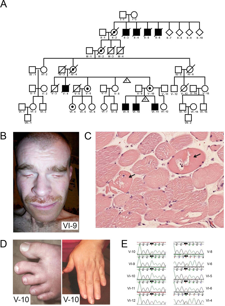
Introduction: Lenz microphthalmia syndrome (LMS) is a genetically heterogeneous X-linked disorder characterised by microphthalmia/anophthalmia, skeletal abnormalities, genitourinary malformations, and anomalies of the digits, ears, and teeth. Intellectual disability and seizure disorders are seen in about 60% of affected males. To date, no gene has been identified for LMS in the microphthalmia syndrome 1 locus (MCOPS1). In this study, we aim to find the disease-causing gene for this condition.

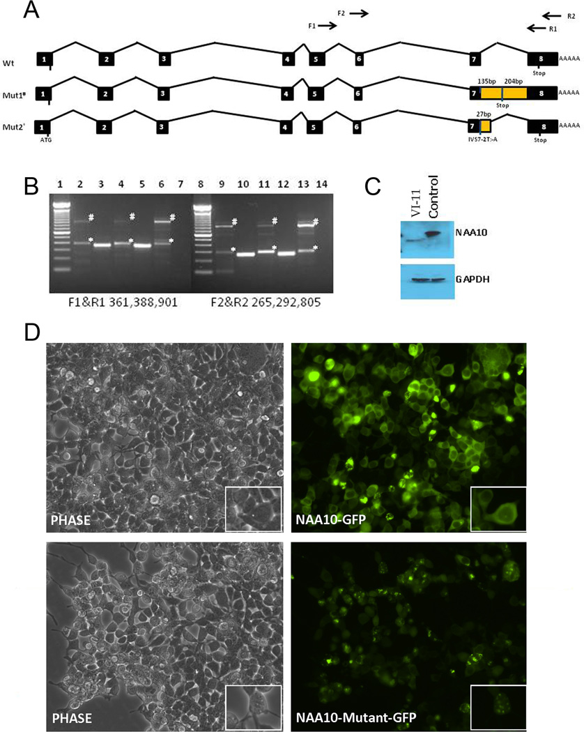
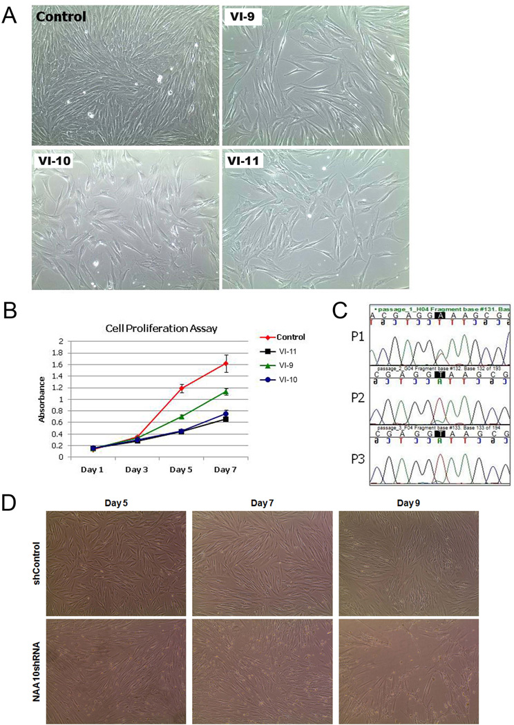
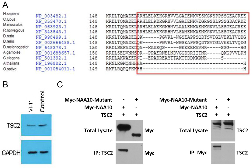
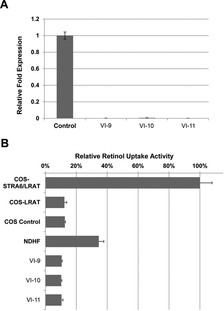
Methods and results: Using exome sequencing in a family with three affected brothers, we identified a mutation in the intron 7 splice donor site (c.471+2T→A) of the N-acetyltransferase NAA10 gene. NAA10 has been previously shown to be mutated in patients with Ogden syndrome, which is clinically distinct from LMS. Linkage studies for this family mapped the disease locus to Xq27-Xq28, which was consistent with the locus of NAA10. The mutation co-segregated with the phenotype and cDNA analysis showed aberrant transcripts. Patient fibroblasts lacked expression of full length NAA10 protein and displayed cell proliferation defects. Expression array studies showed significant dysregulation of genes associated with genetic forms of anophthalmia such as BMP4, STRA6, and downstream targets of BCOR and the canonical WNT pathway. In particular, STRA6 is a retinol binding protein receptor that mediates cellular uptake of retinol/vitamin A and plays a major role in regulating the retinoic acid signalling pathway. A retinol uptake assay showed that retinol uptake was decreased in patient cells.

Conclusions: We conclude that the NAA10 mutation is the cause of LMS in this family, likely through the dysregulation of the retinoic acid signalling pathway.

Keywords: Clinical Genetics; Developmental; Genome-Wide; Lenz Microphthalmia Syndrome; NAA10.


Figures

Figure 1
Figure 1. Pedigree of family affected with Lenz microphthalmia syndrome (LMS), supporting clinical features, and confirmation by Sanger sequencing
(A) Pedigree of LMS affected family. Further experimentation and analyses focused on the three living affected brothers (VI-9, VI-10, VI-11), one obligate heterozygote (V-10), one heterozygote identified previously by linkage (V-6) and her daughter (VI-4). Analysis of family pedigree was consistent with sex-linked inheritance. (B) Bilateral anophthalmia is an example of a dysmorphic feature present in patients affected by LMS (VI-9). (C) Haematoxylin and eosin staining of skeletal muscle showing neuropathic degeneration (arrow). (D) Dysmorphic features such as cutaneous syndactyly between the second and third toes and short terminal phalanges are present in a heterozygote (V-10). (E) Sanger sequencing of NAA10 gene (genomic DNA) shows the c.471+2T→A mutation present in all three affected males (VI-9, VI-10, VI-11), their obligate heterozygote mother (V-10), as well as an aunt who was previously suspected as a heterozygote by linkage (V-6) and one of her daughters (VI-4).
Figure 2
Figure 2. Transcript analysis of mutant NAA10 and implications of exon 8 truncation
(A) Intron/exon structure of NAA10 and variant transcripts present in patient VI-11 with Lenz microphthalmia syndrome. (B) RT-PCR analysis of NAA10 transcripts in control and patients VI-9 and VI-11. Total RNA from patient VI-9 blood, patient VI-11 fibroblasts and skeletal muscle and control fibroblasts was isolated and used for cDNA synthesis and amplification of two NAA10 cDNA amplicons. No NAA10 wild-type band was present in any of the affected male patient samples. Mutant splice variant 1 and 2 are indicated by # and *, respectively. Lanes 2–6: Amplicon F1&R1 (exon 5 and exon 8); lane 1, 100 bp ladder; lane 2, VI-9 blood; lane 3, control blood; lane 4, patient VI-11 muscle; lane 5, control fibroblasts; lane 6, patient VI-11 fibroblasts; lane 7, negative control, no template; lane 8, 100 bp ladder. Lanes 9–14: Amplicon F2&R2 (exon 5/6 junction and exon 8); lane 9, VI-9 blood; lane 10, control blood; lane 11, patient VI-11 muscle; lane 12, control fibroblasts; lane 13, patient VI-11 fibroblasts; lane 14, negative control, no template. (C) Western blot analysis of NAA10 expression in normal and patient VI-11 fibroblast cells. Left lane, NAA10 from patient VI-11; right lane, control fibroblast. #, Splice mutant variant 1. Blots were stripped and re-probed with mouse anti-GAPDH antibody to verify equal loading as shown in the lower panel. (D) Immunofluorescence analysis of wild-type and mutant-NAA10 cellular localisation. NAA10-GFP and mutant-NAA10-GFP were over-expressed in 293T cells. Mutant NAA10-GFP aggregates in the cytoplasm. Top panel, left, phase contrast, right, NAA10-GFP; bottom panel, left, phase contrast, right, NAA10-mutant-GFP.
Figure 3
Figure 3. NAA10 mutation c.471+2T→A causes growth deficiency
(A) Cell proliferation of patient VI-9, VI-10, and VI-11 fibroblasts as compared to control fibroblasts. An equal number of patient and fibroblast cells were plated and phase images were obtained on day 5 of culture; n=3. (B) MTT assay. Control and patient VI-9, VI-10, and VI-11 fibroblast cells were used for the MTT (3-[4,5-dimethylthiazol-2-yl]-2,5-diphenyltetrazolium bromide) assay. 1×10⁵ cells were plated for each cell type, in triplicate, and the assay was performed on days 1, 3, 5, and 7; n=3. (C) Cell growth competition assay. Equal number of control and patient fibroblast cells were mixed and plated in a single well of a 6-well plate. Cells were collected at every passage, for three passages, and DNA purified. The percentage of wild-type or patient cells present at each passage was estimated by chromatogram analysis at the c.471+2T→A mutation site. (D) NAA10 knockdown in normal human fibroblast cells affects cell proliferation. Normal human fibroblast cells were transduced with either shControl or NAA10 shRNA virus. Phase images were obtained on days 5, 7, and 9 after selection with puromycin. By day 9, NAA10 knockdown fibroblast cells became unstable and began to die while shControl fibroblast cells continued to proliferate.
Figure 4
Figure 4. Mutation of NAA10 abolishes the interaction with TSC2
(A). Conservation of amino acid sequence encoded by exon 8 of NAA10. Alignment was done using the Interactive Structure based Sequences Alignment Program Strap (http://3d-alignment.eu/). (B) Mutant NAA10 fails to precipitate TSC2. Western blot analysis of TSC2 expression in a male patient with Lenz microphthalmia syndrome. Left lane, TSC2 from patient VI-11; right lane, TSC2 from control fibroblast. Blots were stripped and re-probed with mouse anti-GAPDH antibody to verify equal loading as shown in the lower panel. (C) Co-immunoprecipitation of NAA10 and TSC2. Immunoprecipitation assays were performed using either anti-TSC2 or anti-Myc antibodies followed by western blot analysis. Mutant NAA10 failed to precipitate TSC2 and vice versa.
Figure 5
Figure 5. Retinol uptake is dramatically affected in patient fibroblast cells
(A) Quantitative real-time PCR analysis of STRA6 expression. RNA was isolated from control and patient VI-9, VI-10, and VI-11 fibroblast cells. STRA6 is significantly down-regulated in patient fibroblast cells. (B) Retinol uptake in control and patient VI-9, VI-10, and VI-11 fibroblast cells. COS untransfected and COS eGFP/LRAT were used as negative controls. COS bSTRA6/LRAT was used as a positive control for the assay.

References

    1. Traboulsi EI, Lenz W, Gonzales-Ramos M, Siegel J, Macrae WG, Maumenee IH. The Lenz microphthalmia syndrome. Am J Ophthalmol. 1988;105:40–45. - PubMed
    1. Bardakjian TM, Schneider AS, Ng D, Johnston JJ, Biesecker LG. Association of a de novo 16q copy number variant with a phenotype that overlaps with Lenz microphthalmia and Townes-Brocks syndromes. BMC Med Genet. 2009;10:137. - PMC - PubMed
    1. Graham CA, Redmond RM, Nevin NC. X-linked clinical anophthalmos. Localization of the gene to Xq27-Xq28. Ophthalmic Paediatr Genet. 1991;12:43–48. - PubMed
    1. Forrester S, Kovach MJ, Reynolds NM, Urban R, Kimonis V. Manifestations in four males with and an obligate carrier of the Lenz microphthalmia syndrome. Am J Med Genet. 2001;98:92–100. - PubMed
    1. Ng D, Hadley DW, Tifft CJ, Biesecker LG. Genetic heterogeneity of syndromic X-linked recessive microphthalmia-anophthalmia: is Lenz microphthalmia a single disorder? Am J Med Genet. 2002;110:308–314. - PubMed

Publication types

Supplementary concepts