

Case Reports
doi: 10.1007/s12663-010-0148-y.
Epub 2011 Feb 4.
Sequestrating giant complex odontoma: a case report and review of the literature
Affiliations
- PMID: 24431893
- PMCID: PMC3847030
- DOI: 10.1007/s12663-010-0148-y
Item in Clipboard
Case Reports
Sequestrating giant complex odontoma: a case report and review of the literature
J Maxillofac Oral Surg.
2013 Dec.
Abstract
Odontomas are the most common benign tumours of odontogenic origin. Due to their hamartomatous nature, they are usually asymptomatic but can cause impaction of one or more teeth. They consist microscopically of all the tissue types found in a developed tooth. We present a case of a large sequestrating complex odontoma resulting in facial asymmetry, cellulitis, pain and partial loss of function. This case has significance, as odontomas of this large size have rarely been reported.
Keywords: Complex odontoma; Hamartoma; Odontogenic; Radiographic features; Sequestration.
Figures
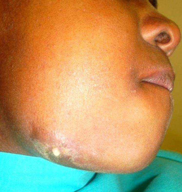
Swelling with draining sinus and sub-mandibular cellulitis at the lower right mandibular border
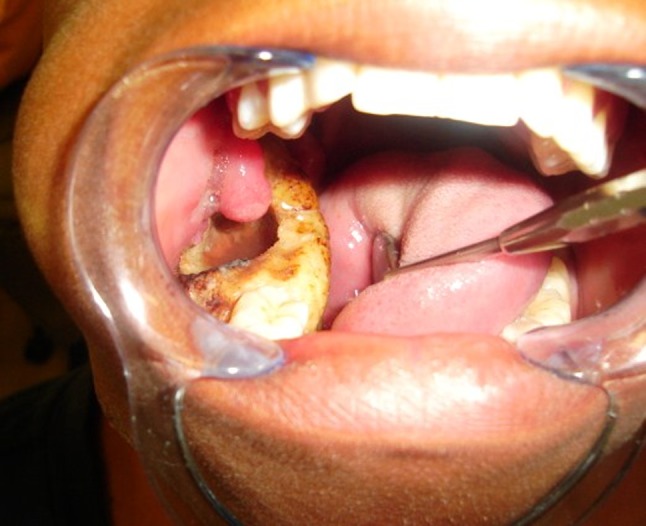
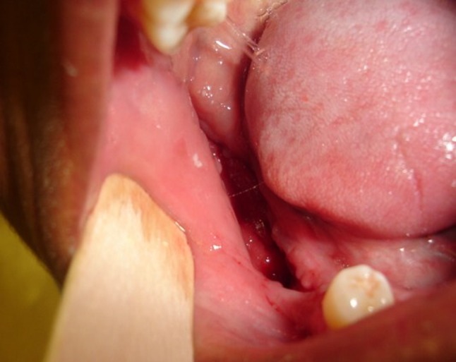
Intraoral view of the lesion
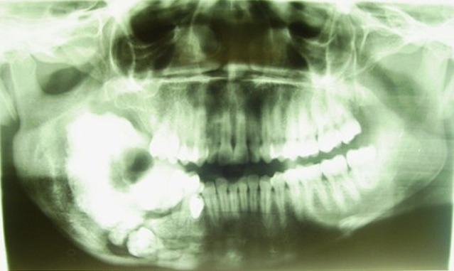
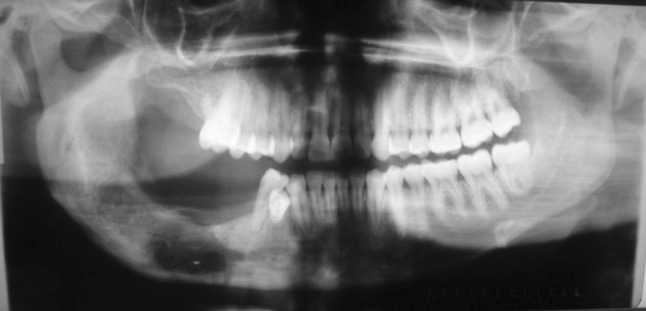
Pre-operative panoramic radiograph 2008; the lesion has grown since 2004 and shows clearly a well-defined radiolucent rim which is the result of overlying infection

Panoramic radiograph (2004); the extension of the radiopaque lesion into the ramus and the impacted first molar above the inferior border of the mandible is clearly visible. The lesion is surrounded by a radiolucent border, which seems to be more pronounced in the area of the ramus
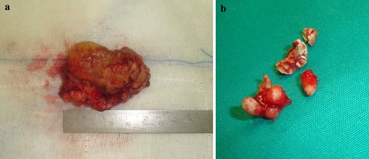
a Specimen sample of lesion. b Fractured impacted tooth after removal
Panoramic radiograph after removal of BIPP
Intra-oral view 3 months later—note the healing by secondary intention

Micrograph shows a haphazard mixture of odontogenic hard tissue. Extensive necrosis and inflammation was present. (Histopathology, courtesy: Prof E. J. Raubenheimer)
References
-
- Cildir SK, Sencift K, Olgac V, Sandalli N (2005) Delayed eruption of a mandibular primary cuspid associated with compound odontoma. J Contemp Dent Pract 15;6(4):152–159 - PubMed
-
- Kramer IRH, Pindborg JJ, Shear M. Histological typing of odontogenic tumours. World Health Organization. International histological classification of tumours. 2. Berlin: Springer; 1992. pp. 16–21.
-
- Noffke CEE, Chabikuli NJ, Nzima N (2005) Impaired tooth eruption: a review. SADJ 60(10):422, 424–425 - PubMed
-
- Serra-Serra G, Berini-Aytes L, Gay-Escoda C. Erupted odontomas: a report of three cases and review of the literature. Med Oral Patol Oral Cir Bucal. 2009;14(6):E299–E303. - PubMed
Publication types
LinkOut - more resources
Full Text Sources