

Cerebral microbleeds: a review of clinical, genetic, and neuroimaging associations
- PMID: 24432010
- PMCID: PMC3881231
- DOI: 10.3389/fneur.2013.00205
Cerebral microbleeds: a review of clinical, genetic, and neuroimaging associations
Abstract
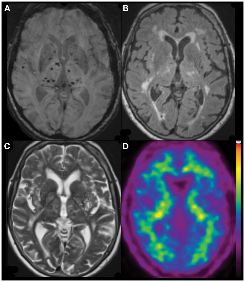
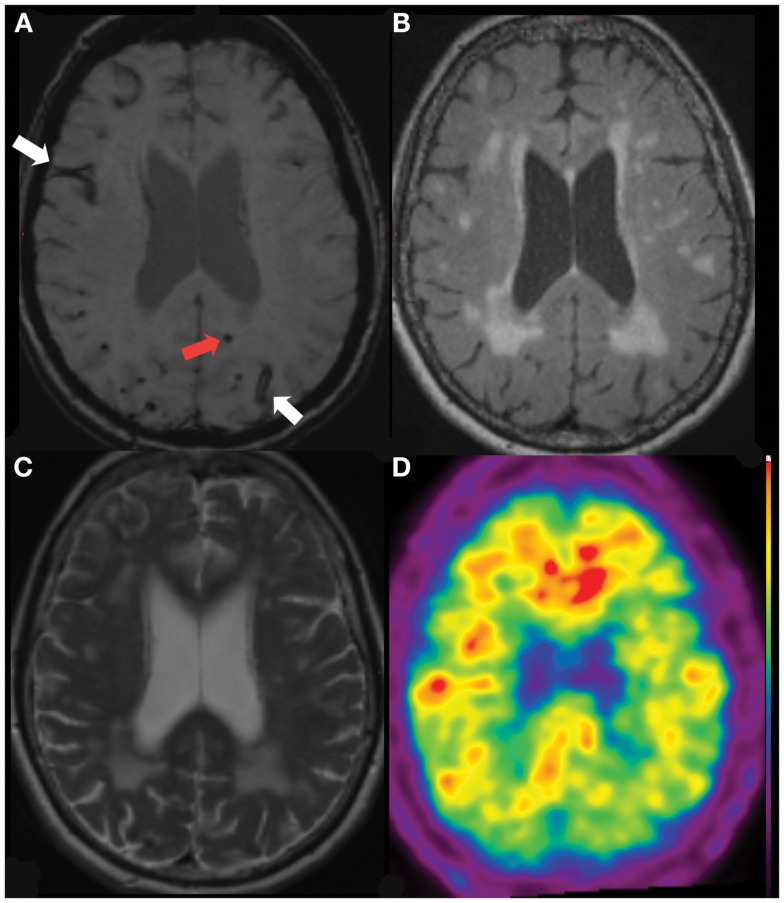
Cerebral microbleeds (microbleeds) are small, punctuate hypointense lesions seen in T2* Gradient-Recall Echo (GRE) and Susceptibility-Weighted (SWI) Magnetic Resonance Imaging (MRI) sequences, corresponding to areas of hemosiderin breakdown products from prior microscopic hemorrhages. They occur in the setting of impaired small vessel integrity, commonly due to either hypertensive vasculopathy or cerebral amyloid angiopathy. Microbleeds are more prevalent in individuals with Alzheimer's disease (AD) dementia and in those with both ischemic and hemorrhagic stroke. However they are also found in asymptomatic individuals, with increasing prevalence with age, particularly in carriers of the Apolipoprotein (APOE) ε4 allele. Other neuroimaging findings that have been linked with microbleeds include lacunar infarcts and white matter hyperintensities on MRI, and increased cerebral β-amyloid burden using (11)C-PiB Positron Emission Tomography. The presence of microbleeds has been suggested to confer increased risk of incident intracerebral hemorrhage - particularly in the setting of anticoagulation - and of complications of immunotherapy for AD. Prospective data regarding the natural history and sequelae of microbleeds are currently limited, however there is a growing evidence base that will serve to inform clinical decision-making in the future.
Keywords: Alzheimer’s disease; MRI imaging; amyloid imaging; cerebral amyloid angiopathy; intracerebral hemorrhage; microbleeds; positron-emission tomography; stroke.
Figures
References
-
- Matsukawa H, Shinoda M, Fujii M, Takahashi O, Yamamoto D, Murakata A, et al. Factors associated with lobar vs. non-lobar intracerebral hemorrhage. Acta Neurol Scand (2011) [cited 2012 Feb 29]. Available from: http://www.ncbi.nlm.nih.gov/pubmed/22067041 - PubMed
Publication types
LinkOut - more resources
Full Text Sources
Other Literature Sources
Miscellaneous