. 2014 Jan 6:4:502.
doi: 10.3389/fimmu.2013.00502.

Anti-CD20 as the B-Cell Targeting Agent in a Combined Therapy to Modulate Anti-Factor VIII Immune Responses in Hemophilia a Inhibitor Mice

Affiliations

Anti-CD20 as the B-Cell Targeting Agent in a Combined Therapy to Modulate Anti-Factor VIII Immune Responses in Hemophilia a Inhibitor Mice

Chao Lien Liu et al. Front Immunol. .

Abstract

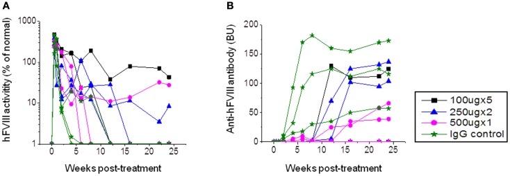
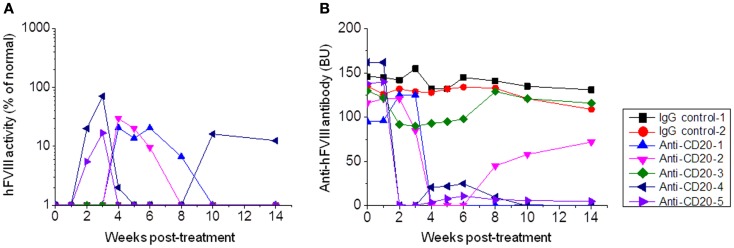
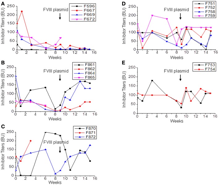
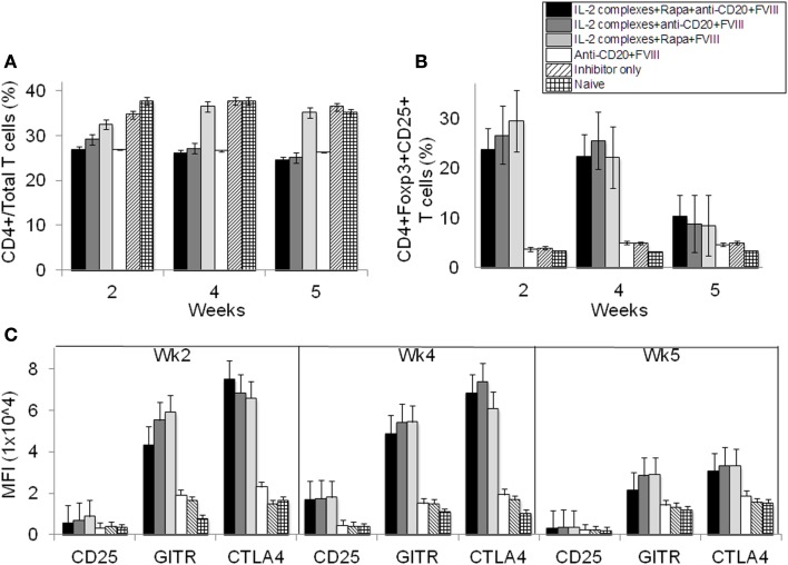
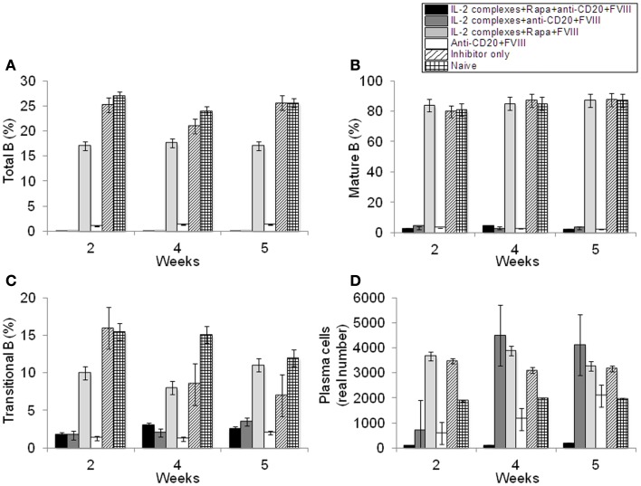
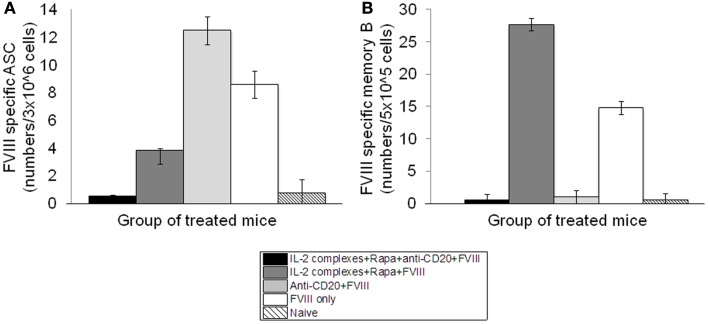
Neutralizing antibody formation against transgene products can represent a major complication following gene therapy with treatment of genetic diseases, such as hemophilia A. Although successful approaches have been developed to prevent the formation of anti-factor VIII (FVIII) antibodies, innovative strategies to overcome pre-existing anti-FVIII immune responses in FVIII-primed subjects are still lacking. Anti-FVIII neutralizing antibodies circulate for long periods in part due to persistence of memory B-cells. Anti-CD20 targets a variety of B-cells (pre-B-cells to mature/memory cells); therefore, we investigated the impact of B-cell depletion on anti-FVIII immune responses in hemophilia A mice using anti-CD20 combined with regulatory T (Treg) cell expansion using IL-2/IL-2mAb complexes plus rapamycin. We found that anti-CD20 alone can partially modulate anti-FVIII immune responses in both unprimed and FVIII-primed hemophilia A mice. Moreover, in mice treated with anti-CD20+IL-2/IL-2mAb complexes+rapamycin+FVIII, anti-FVIII antibody titers were significantly reduced in comparison to mice treated with regimens targeting only B or T cells. In addition, titers remained low after a second challenge with FVIII plasmid. Treg cells and activation markers were transiently and significantly increased in the groups treated with IL-2/IL-2mAb complexes; however, significant B-cell depletion was obtained in anti-CD20-treated groups. Importantly, both FVIII-specific antibody-secreting cells and memory B-cells were significantly reduced in mice treated with combination therapy. This study demonstrates that a combination regimen is highly promising as a treatment option for modulating anti-FVIII antibodies and facilitating induction of long-term tolerance to FVIII in hemophilia A mice.

Keywords: B-cell depletion; anti-CD20; factor VIII; hemophilia; immunomodulation; tolerance induction.


Figures

Figure 1
Figure 1
Factor VIII gene expression and anti-FVIII antibody formation after FVIII plasmid+anti-CD20 treatment in hemophilia A mice. Four groups of hemophilia A mice were treated with FVIII plasmid (50 μg/treatment/mouse) at day 0 and i.v. injection of anti-CD20 at various doses and schedules as listed in the following: group 1: 100 μg/treatment/mouse, injected at days −2, 0, 3, 6, and 9. Group 2: 250 μg/treatment/mouse, injected at days 0 and 14. Group 3: 500 μg/treatment/mouse, injected at day 0. Group 4: control rat IgG, 250 μg/treatment/mouse, injected at days 0 and 14. Peripheral blood samples were collected at different time points to examine FVIII activities (A) and inhibitor titers (B). Each symbol represents data obtained from an individual mouse. Data shown is representative of two independent experiments.
Figure 2
Figure 2
Factor VIII gene expression and anti-FVIII antibody titers following anti-CD20 treatment in FVIII plasmid-primed hemophilia A mice with pre-existing inhibitors. Mice were primed with FVIII plasmid to induce high-titer inhibitory antibodies at 8 weeks before anti-CD20 treatment. The inhibitor mice were then treated with anti-CD20. Group 1: control rat IgG, 250 μg/treatment/mouse, injected at days −7, −4, and 0. Group 2: anti-CD20, 250 μg/treatment/mouse, injected at days −7, −4, and 0. Peripheral blood samples were collected at different time points to evaluate FVIII activities (A) and inhibitor titers (B). Each symbol represents data obtained from an individual mouse. Data shown is representative of two independent experiments.
Figure 3
Figure 3
Immunomodulation with separate or combined therapy by IL-2/IL-2mAb complexes, rapamycin, and anti-CD20 in FVIII plasmid-primed hemophilia A mice with pre-existing inhibitors. Four groups of hemophilia A inhibitor mice were treated separately with different combined regimens: (A) IL-2/IL-2mAb complexes+rapamycin+ anti-CD20+FVIII injection (n = 4, group 1), (B) IL-2/IL-2mAb complexes+anti-CD20+FVIII (n = 4, group 2), (C) IL-2/IL-2mAb complexes+rapamycin+FVIII injection (n = 3, group 3), (D) anti-CD20+FVIII injection (n = 4, group 4), and (E) Control inhibitor mice (n = 2, group 5). Each experimental group was treated with indicated immunomodulation regimen weekly for 4 weeks. Anti-FVIII antibody titers were assessed by Bethesda assay over time. Each symbol represents data obtained from an individual mouse. Data shown is representative of three independent experiments.
Figure 4
Figure 4
Effects of immunomodulation on CD4+ T cells, CD4+CD25+Foxp3+ Treg cells and Tregs activation markers in peripheral blood of treated hemophilia A inhibitor mice over time. Lymphocytes were isolated from the blood of naive (light grid), inhibitor only (light slant), anti-CD20+FVIII (white), IL-2/IL-2mAb complexes+rapamycin+FVIII (light gray), IL-2/IL-2mAb complexes+anti-CD20+FVIII (dark gray), and IL-2/IL-2mAb complexes+rapamycin+anti-CD20+FVIII (black) treated mice. (A) CD4+ in total T cells and (B) CD4+CD25+Foxp3+ in CD4+ T cells were stained and analyzed by flow cytometry during the treatment. (C) Blood cells were also stained and analyzed for Treg cells markers: CD25, GITR, and CTLA-4. Data shown are median fluorescence intensity (MFI) values of the three activation markers. Data shown is representative of two independent experiments.
Figure 5
Figure 5
Effects of immunomodulation on total B, mature B, transitional B, and plasma B-cells in peripheral blood of each mouse group. Lymphocytes were isolated from the blood of naive (light grid), Inhibitor only (light slant), anti-CD20+FVIII (white), IL-2/IL-2mAb complexes+rapamycin+FVIII (light gray) IL-2/IL-2mAb complexes+anti-CD20+FVIII (dark gray), and IL-2/IL-2mAb complexes+rapamycin+anti-CD20+FVIII (black) treated mice. (A) B220+ B-cells, (B) IgM+IgDhi B-cells, (C) transitional B-cells, and (D) plasma cells were stained and analyzed by flow cytometry during the treatment period (week 2, 4, and 5). Data shown are cell percentages (A–C) and real numbers (D) of the B-cell populations. Data shown is representative of two independent experiments.
Figure 6
Figure 6
Depletion of FVIII-specific ASCs (Antibody-Secreting Cells) and memory B-cells in the inhibitor mice treated with IL-2/IL-2mAb complexes plus rapamycin and anti-CD20. Cells were isolated by MACS from spleens of naive (light slant), FVIII plasmid only (white), anti-CD20+FVIII (light gray), IL-2/IL-2mAb complexes+rapamycin+FVIII (dark gray), and IL-2/IL-2mAb complexes+rapamycin+anti-CD20+FVIII (black) treated mice (n = 2, each group) 2 weeks after treatment. (A) 3 × 106 cells were used to detect FVIII-specific ASC cells by ELISPOT assay. (B) 5 × 105 non-plasma cells were cultured and stimulated with FVIII at 10 U/ml for 6-days, and memory B-cells were detected by ELISPOT assay. Data shown are mean of spot numbers for each treated group (n = 2).

Similar articles

Cited by

References

    1. Darby SC, Keeling DM, Spooner RJ, Wan Kan S, Giangrande PL, Collins PW, et al. The incidence of factor VIII and factor IX inhibitors in the hemophilia population of the UK and their effect on subsequent mortality, 1977–99. J Thromb Haemost (2004) 2(7):1047–5410.1046/j.1538-7836.2004.00710.x - DOI - PubMed
    1. Ehrenforth S, Kreuz W, Scharrer I, Linde R, Funk M, Güngör T, et al. Incidence of development of factor VIII and factor IX inhibitors in haemophiliacs. Lancet (1992) 339(8793):594–810.1016/0140-6736(92)90874-3 - DOI - PubMed
    1. Grillo-Lopez AJ, Hedrick E, Rashford M, Benyunes M. Rituximab: ongoing and future clinical development. Semin Oncol (2002) 29(1 Suppl 2):105–1210.1053/sonc.2002.30145 - DOI - PubMed
    1. Bour-Jordan H, Bluestone JA. B cell depletion: a novel therapy for autoimmune diabetes? J Clin Invest (2007) 117(12):3642–510.1172/JCI34236 - DOI - PMC - PubMed
    1. Boye J, Elter T, Engert A. An overview of the current clinical use of the anti-CD20 monoclonal antibody rituximab. Ann Oncol (2003) 14(4):520–3510.1093/annonc/mdg175 - DOI - PubMed