

A brief overview of 100 years of history of surgical treatment for adolescent idiopathic scoliosis
- PMID: 24432060
- PMCID: PMC3566253
- DOI: 10.1007/s11832-012-0466-3
A brief overview of 100 years of history of surgical treatment for adolescent idiopathic scoliosis
Abstract
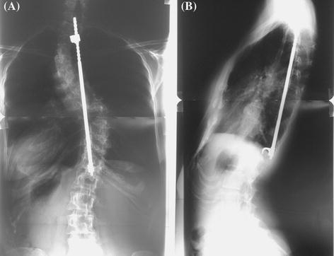
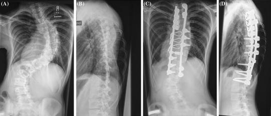
The history of surgical correction for adolescent idiopathic scoliosis reaches back about 100 years: the natural course of progressive, crippling and sometimes even life-threatening deformities which could not be controlled by external means called for effectual, invasive procedures. Hibbs 1911 aimed at halting progression by long, uninstrumented fusions. However, the lack of true correction, long rehabilitation times, high pseudarthrosis and infection rates, and a fusion mass which bent further once exposed to gravity again were not satisfying. The transition from slowing progression to halting progression and truly correcting the deformity lasted almost another half a century: Paul Harrington, confronted with many scoliotic polio patients, successfully introduced a hook-rod system for concave-distraction and convex-compression at the end of the 1950s. Many implant failures, a still-considerable pseudarthrosis rate, flattening of the sagittal profile and the lack of true three-dimensional (3D) correction were the shortcomings. In the 1970s the Frenchmen Cotrel and Dubousset took scoliosis surgery to the next level by introducing a versatile hook system and curve-pattern-adapted correction modes. The basics of the so-called derotation-manoeuvre consists in strategic distribution of the anchors along the curve, bending the rod accordingly, and rotating it back into the sagittal plane. The overall correction, stability and the fusion rates improved significantly. However, the effect on the sagittal and transverse plane were still limited. Lately, a better biomechanical understanding and bilateral, polysegmental strong three-column fixation with pedicle screw has become the benchmark method: in conjunction with posterior release techniques, osteotomies or even vertebral column resections for severe cases, it allows better 3D control (vertebral column manipulation), faster rehabilitation and better patient satisfaction.
Keywords: Adolescent; History; Idiopathic scoliosis; Surgery.
Figures
References
-
- Zielke K. The so-called Risser plaster technic. Z Orthop Ihre Grenzgeb. 1971;109:341–344. - PubMed
-
- Harrington PR. Treatment of scoliosis. Correction and internal fixation by spine instrumentation. J Bone Joint Surg Am. 1962;44-A:591–610. - PubMed
-
- Harrington PR. Present status of spine instrumentation in scoliosis. Am J Orthop. 1963;5:228–231. - PubMed
Publication types
LinkOut - more resources
Full Text Sources
Other Literature Sources
